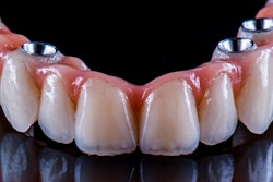
Two-step techniques may improve the trueness of complete arch trial restorations compared to those that only use a one-step method, and more supporting teeth may reduce deviations. The study was published recently in BMC Oral Health.
Furthermore, two-step mock-up techniques with different stop placements may enhance the accuracy of complete arch trial restorations, the authors wrote.
“This study demonstrates that two-step mock-up methods significantly enhance the trueness of complete-arch trial restorations compared to conventional one-step approaches, with trueness progressively improving as the number of supporting teeth increases,” wrote the authors, led by Chunxiao Jin of Wuhan University in China (BMC Oral Health, October 10, 2025, Vol. 25, 1588).
The study’s intent was to evaluate how accurately complete arch trial restorations could be fabricated using a two-step mock-up method with different stop distributions compared to a conventional one-step technique. Researchers designed complete arch digital diagnostic waxing on a worn maxillary cast from a patient with severely worn dentition, they wrote.
The composite waxing cast was used to fabricate one-step trial restorations (Group 1) and as the base for the second step of the two-step mock-up process (Groups 2 to 5) with varying retained teeth. For each two-step method, partial restorations were first created using a silicone index and then completed with bis-acrylic resin injected into the index, seated on the cast under consistent pressure, and fully cured before removal.
Regarding trueness, significant differences were observed among the five groups (F-value (4,20) = 92.61; p < 0.001). Group 1 had the largest mean root mean square deviation (0.31 ± 0.02 mm), while Group 5 (alternating teeth) had the smallest (0.15 ± 0.01 mm), showing no significant difference from Group 4 (0.17 ± 0.01 mm), they wrote.
Tooth-level 3D-analysis showed the greatest deviations at the premolars (0.34 ± 0.07 mm) and canines (0.60 ± 0.09 mm) in Group 1, both significantly higher than Groups 4 and 5 (< 0.17 mm, all p < 0.001). Occlusal surface 2D-analysis found significant differences (all p < 0.001), with Group 1 showing a 0.58 ± 0.12 mm deviation at the premolars versus 0.16 ± 0.09 mm in Group 5. Overall, trueness improved as the number of supporting teeth increased, reducing deviations in nearby teeth.
This study, however, had limitations. The study’s generalizability may be limited because it relied on a single silicone material and one provisional dental material, the authors added.
“Further research is needed to evaluate the two-step mock-up method’s applicability in varied intraoral scenarios and across different restoration types, as well as its performance with other materials in clinical practice,” Jin and colleagues wrote.


