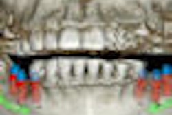
The majority of postgraduate orthodontic programs in the U.S. and Canada has access to and regularly use cone-beam CT (CBCT) as part of their hands-on curriculum, according to a study in the Journal of Dental Education (January 2011, Vol. 75:1, pp. 98-106).
Researchers from A.T. Still University sent an anonymous electronic survey to the program director/chair of all 69 U.S. and Canadian postgraduate orthodontic programs. Of these, 36 (52.2%) of the programs responded.
Overall, 83% of these programs reported having access to a cone-beam CT scanner, while 73.3%. The vast majority (82%) used cone-beam CT mainly for specific diagnostic purposes -- such as evaluating impacted/supernumerary teeth, craniofacial anomalies, and temporomandibular joint (TMJ) disorders -- while 18% used it as a diagnostic tool for every patient. Orthodontic residents received a combination of didactic and hands-on training (59%) or solely didactic training (32%).
Operation of the scanner was the responsibility of radiology technologists (54%), both radiology technologists and orthodontic residents (32%), and orthodontic residents alone (14%). Interpretation of the results was the responsibility of a radiologist in 59% of programs, while residents were responsible for reading and referring abnormal findings in 32% of the programs.
Copyright © 2011 DrBicuspid.com
