A woman presented with an extremely rare case of bilateral fusion of her permanent lower jaw lateral incisors and canines, which resulted in crowding and made it difficult to clean her teeth. The case report was published on January 11 in Clinical Case Reports.
It is believed to be the first reported case of this particular type of bilateral fusion, the authors wrote.
“This case highlights the importance of a thorough diagnostic approach, including detailed medical and dental history, comprehensive clinical examination, and radiographic assessment,” wrote the authors, led by Siddharthan Selvaraj of the University of Puthisastra in Cambodia.
A 35-year-old woman with a developmental anomaly
The woman went to a dental clinic, complaining of bleeding gums. Her medical history was unremarkable, including no history of dental trauma, according to the case report.
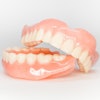
An oral exam showed that the patient had bilateral fusion of the right and left lower jaw permanent lateral incisors and canines, which formed a single large crown with a developmental groove on the labial and lingual aspects and extending into the cervical area. She had some crowding of her incisors, as well as some gum inflammation. Additionally, the woman had no other developmental abnormalities, the authors wrote.
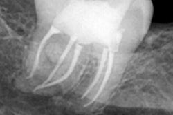
 (A) Intraoral photograph showing bilaterally fused mandibular lateral incisors and canines with visible developmental grooves on the labial surfaces and mild tooth surface loss in relation to teeth #42 and #43. (B,C) Intraoral periapical radiographs demonstrating bilateral fusion of the mandibular lateral incisors and canines, with two distinct coronal pulp horns converging into a single pulp chamber at the cervical level. (D) Mandibular occlusal radiograph confirming bilateral fusion of the lateral incisors and canines.Images and captions courtesy of Selvaraj et al. Licensed under CC BY 4.0 International.
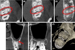
Periapical x-rays revealed that her fused teeth showed a single root with a single canal, and this was confirmed on cone-beam computed tomography (CBCT). Furthermore, the fused teeth looked very similar on imaging.
“Symmetrical fusion likely resulted from the simultaneous contact between developing tooth buds on both sides of the lower jaw,” they wrote. “… Each fused tooth exhibited a single root, suggesting that the fusion occurred before crown calcification, resulting in the complete fusion of both crown and root structures.”
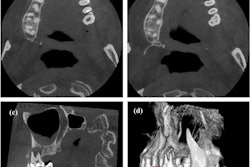
 (A-C) show axial CBCT images at the cervical, middle, and apical thirds of the root, respectively, of the fused right and left lateral incisors and canines exhibiting a single pulp chamber and a single root. (D, E) show sagittal CBCT sections demonstrating similar crown and root morphology of the fused lateral incisor and canine on both sides. Minor image noise was noted, partially obscuring fine internal details of the root canals; nevertheless, the diagnostic interpretation remained unaffected.
Although the woman had noticeable crowding and difficulty maintaining effective plaque control, she declined orthodontic correction due to cost. The woman was told to visit the dentist every six months to evaluate plaque control, gum health, and occlusal stability, the authors wrote.
What to know about fusion
Fusion is an unusual developmental anomaly that is defined by the meeting of two adjacent teeth during the morphodifferentiation stage of tooth formation, which is when the final crown shape is determined.
The pervasiveness of fusion occurring in primary teeth ranges from 0.4% to 0.9%, and it’s about 0.2% in permanent teeth. While unilateral fusion is more commonly seen in primary teeth over permanent teeth, bilateral fusion in permanent teeth is very rare, with a reported prevalence of only 0.05%, the authors wrote.
Moreover, fusion in primary teeth has been linked to a heightened risk of tooth decay, and it can also affect permanent teeth. Therefore, detecting this dental anomaly early is crucial to prevent possible negative effects on the permanent dentition. In addition to the increased risk of tooth decay, fusion in permanent teeth leads to problems, including crowding and abnormal occlusion.
“This rare clinical case gives the importance of morphological and radiological investigation ensuring optimal aesthetic and functional outcome,” Selvaraj and colleagues wrote.