Japanese researchers have found that several PET/CT parameters -- in particular, a ring-shaped area of FDG uptake -- are reliable predictors of survival for patients with head and neck squamous cell carcinoma (HNSCC). Results of their study were published in the April issue of the American Journal of Roentgenology.
The retrospective study evaluated four parameters of FDG uptake -- maximum standardized uptake value (SUVmax), metabolic tumor volume, total lesion glycolysis, and FDG uptake pattern -- and found that all provided valuable information regarding patient survival.
Of the parameters, the strongest predictor was a qualitative one, ring-shaped FDG uptake, according to lead author Dr. Sho Koyasu, from the department of diagnostic imaging and nuclear at medicine at Kyoto University Graduate School of Medicine, and colleagues (AJR, April 2014, Vol. 202:4, pp. 851-858).
Staging head and neck cancer
TNM stage is a primary prognostic tool for cancer and is often used to guide treatment decisions, the authors wrote. T refers to the primary tumor site, N describes regional lymph node involvement, and M denotes whether distant metastasis has occurred. However, TNM staging does not always provide satisfactory results because each tumor stage encompasses a range of population and tumor features.
FDG-PET is widely used in oncologic applications and has proved clinically valuable in detecting and staging cancers, as well as for predicting response to neoadjuvant chemotherapy for HNSCC, Koyasu and colleagues noted. SUVmax is a commonly used parameter for FDG-PET, and two new 3D parameters -- metabolic tumor volume (MTV) and total lesion glycolysis (TLG) -- have also shown promise for predicting outcome.
All three parameters are measured quantitatively, but they have shortcomings that may give an incomplete picture of the tumor. The researchers speculated that a qualitative parameter, FDG uptake pattern, could also be useful for patients with HNSCC.
To test the theory, the group conducted a retrospective study that included 108 patients with confirmed oral, oropharyngeal, hypopharyngeal, and laryngeal squamous cell carcinomas. All patients underwent PET/CT (Discovery ST Elite-Performance, GE Healthcare, or Biograph Duo LSO, Siemens Healthcare) between August 2005 and December 2009 prior to treatment. After scanning, 33 people (30%) underwent surgery and 75 (70%) received radiation therapy. Patients with stage III or IV disease received neoadjuvant chemotherapy, and some patients received concomitant chemotherapy.
Koyasu and colleagues evaluated SUVmax on PET/CT for the primary tumor and MTV and TLG summed for all lesions, and they also visually inspected FDG uptake patterns to determine if they were sphere-shaped or ring-shaped. They correlated these parameters with disease-specific survival and disease-free survival to assess their impact on patient outcomes.
The median follow-up period for the surviving patients was 36.4 months (range, 10 weeks to 73 months and four days). There were 89 survivors during the study period, 79 (89%) of whom were followed for more than one year and 71 (80%) of whom were followed for more than two years.
The estimated two-year disease-specific survival rate was 86.5%, and estimated median disease-free survival was 33.4 months. Over the course of the study period, cancer recurrence or related death occurred in 27 (25%) of the 108 patients. The estimated two-year disease-free survival rate was 76.3%.
PET/CT parameters
All of the quantitative FDG parameters significantly predicted patient outcome in terms of disease-specific and disease-free survival. The researchers determined cutoff points for worse outcomes as follows:
- SUVmax greater than 10 g/mL
- Metabolic tumor volume greater than 20 cm3
- Total lesion glycolysis greater than 70 g
For example, more than 90% of patients with SUVmax less than 10 g/mL had an overall survival rate of more than six years. By comparison, only 65% of patients with SUVmax greater than 10 g/mL survived for three years. Patients with SUVmax less than 10 g/mL were nearly six times as likely to survive their disease than those with higher levels (hazard ratio = 5.66).
With respect to metabolic tumor volume, more than 95% of patients with MTV less than 20 cm3 survived for more than six years; meanwhile, approximately 60% of patients with MTV greater than 20 cm3 survived for slightly less than three years. Patients with MTV less than 20 cm3 were nearly nine times as likely to survive than those with higher levels (hazard ratio = 8.89).
For total lesion glycolysis, more than 95% of patients with TLG less than 70 g survived for more than six years, whereas approximately 60% of patients with TLG greater than 70 g survived for slightly less than three years. Patients with TLG less than 70 g were eight times as likely to survive compared to those with higher levels (hazard ratio = 8.38).
Ring of fire
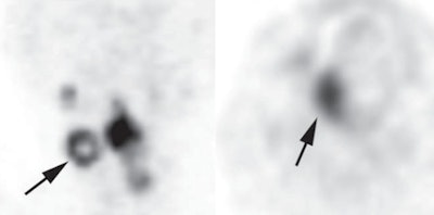
As good as these quantitative factors were, the shape of FDG uptake was better. Patients with sphere-shaped patterns of FDG uptake were almost 19 times more likely to survive than those with ring-shaped patterns (hazard ratio = 18.92).
Of the 108 patients, 14 had ring-shaped patterns and 94 had sphere-shaped patterns. Approximately 90% of those with sphere-shaped patterns survived for more than six years. In comparison, 10% of subjects with ring-shaped patterns survived for more than 36 months.
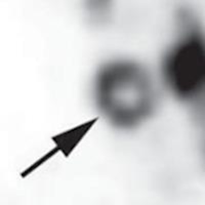
 Left PET image shows typical ring-shaped uptake (arrow) in metastatic lymph node of a 54-year-old woman with hypopharyngeal squamous cell carcinoma. Sphere-shaped uptake shown at right is from a primary lesion in a 55-year-old man with SCC of the oral cavity. Images courtesy of AJR.
"Our findings suggest that the uptake pattern on FDG-PET/CT can provide better prognostic information than conventional quantitative patterns," the authors wrote. This could be because uptake patterns better reflect intratumoral necrosis or heterogeneous morphology, they added.
What's more, the authors believe this could be the first study to demonstrate the significance of the ring-shaped FDG uptake pattern for predicting survival in patients with HNSCC (other studies have looked at the patterns in other types of cancers).
"Furthermore, the uptake pattern could identify patients with poorer outcome in the group with a high MTV, suggesting that it might be used to select therapeutic strategy and that it might add information for predicting a poor prognosis when clinically staging patients with HNSCC," they wrote.
Koyasu and colleagues cited several limitations of the study, including its retrospective nature and the relatively small number of patients. They recommend larger multicenter, prospective, randomized studies to further validate their results.


