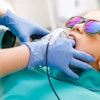
Intraoral ultrasonography provides morphological information that is not accessible with conventional dental x-rays, according to researchers from Paris Descartes University (Clinical Oral Investigations, March 5, 2011).
Although ultrasonography is a noninvasive, inexpensive, and painless diagnostic tool for soft-tissue imaging, this technique is not currently used for oral exploration, the study authors noted.
To determine the diagnostic potential of this technique for dentistry, they developed a 25-MHz high-frequency ultrasound probe designed especially for intraoral applications. Two independent radiologists performed ultrasound examinations on three healthy volunteers. All teeth were explored on the lingual and buccal sides (162 samples) to evaluate the ergonomics of the system and the visualization of anatomic structures.
At the gingivodental junction of the maxillary and mandibular teeth, the device clearly identified the tooth surfaces, the alveolar bone reflection with its surrounding subepithelial connective tissue of the gingival, and the gingival epithelia. The bone level and the thickness of soft tissue around the implant are measurable on the buccal and lingual sides.
"We propose a novel diagnostic tool that explores the biological width and is able to define the thin or thick nature of the gums," the authors wrote. "Moreover, intraoral ultrasonography may help to monitor precancerous lesions."
They recommend large-scale clinical studies to determine whether ultrasonography could be used as a diagnostic tool for daily dental practice.