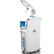
Lasers are traditionally used in dentistry as high-tech surgical tools that can precisely cut both hard and soft tissue and often neatly coagulate in the process.
More recently, laser-based imaging technologies such as quantitative laser fluorescence and optical coherence tomography are being developed specifically for diagnostic applications in dentistry.
Now researchers from Australia and Taiwan are using laser energy to nondestructively analyze the health of human teeth and enable earlier diagnosis of caries lesions. In particular, they evaluated the mineral content of tooth enamel by measuring how the surface of two extracted teeth responded to laser-generated ultrasound.
Their findings appear in the latest issue of Optics Express (August 31, 2009, Vol. 17:8, pp. 15592-15607).
Measuring demineralization
Visual, tactile, and x-ray assessments remain the most common methods used for caries detection in the current clinical setting, said study author David Hsiao-Chuan Wang, Ph.D., a research student at the University of Sydney School of Physics, in an interview with DrBicuspid.com. But all three have their limitations, he noted.
Visual examination is most useful when detecting smooth surface lesions, but is often obscured by plaque or salivary contamination of the tooth structures. Tactile examination can aid the detection of caries in areas that are difficult to see. However, exploration with a sharp instrument can be destructive. X-ray assessment provides visualization of caries in the proximal space between adjacent teeth, but it is sometimes difficult to distinguish between a cavitated and noncavitated lesion.
In addition, all of these conventional clinical techniques are unable to evaluate the level of mineralization, Wang noted.
Surface acoustic waves
Wang's approach utilizes something called an ultrasonic surface acoustic wave (SAW). In their experiments, he and his colleagues sent short-duration laser pulses from an Nd:YAG (neodymium-doped yttrium aluminum garnet) laser onto the surface of two extracted teeth. These pulses induced rapid, microscopic, thermoelastic expansion of the material, which in turn generated a SAW.
The surface acoustic wave probed the entire enamel depth, and its velocity was influenced by all the enamel layers probed. The researchers detected the wave using a noncontact fiber-optic probe connected to an interferometer. The SAW velocity profile allowed the researchers to derive the elasticity response of the measured region as a function of enamel depth.
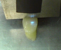 |
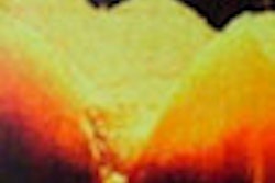
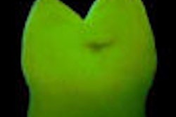
| Researchers evaluate the mineral content of tooth enamel. Image courtesy of David Hsiao-Chuan Wang, Ph.D. |
"The SAW is particularly useful for evaluating elasticity of material because its propagation characteristic is very sensitive to the elastic modulus of the surface material," Wang explained. "The laser pulse energy was low enough such that no visible damage occurred to the enamel."
The SAW technique has significant application in terms of determining the location and the severity of the demineralization sites in the clinical setting, as well as assessing the efficacy of the proposed remineralization treatments for dental research purposes, he added.
"Our technique is noncontact, nondestructive, and, most importantly, it is able to quantify the elasticity variation of dental enamel," Wang said.
Still, a commercial version of the device is likely a few years away. Wang noted that they will need to refine the choice of laser in terms of wavelength, power, and pulse duration to achieve the optimum balance between diagnostic accuracy, safety, and cost. For in vivo use, they also will need to develop a more compact, probably handheld, probe.
"Given the appropriate amount of support, I am confident we will develop something that could be commercialized within two or three years," he said.
Quantitative measurements
This is the first time that anyone has been able to quantitatively measure the elasticity of human teeth nondestructively, according to Donald Coluzzi, D.D.S., editor of the Journal of Laser Dentistry.
"Although dentists conventionally probe (with an explorer) to check the tooth enamel, and that's nondestructive, it's a qualitative result," he wrote in an e-mail to DrBicuspid.com. "So the word 'measure' in terms of quantitative is probably what's being described here."
The importance would be that the tooth hardness (or degree of mineralization) could be accurately determined, he noted.
A compact, handheld probe based on this technology would be a great diagnostic tool, he stated.
"There's emerging technology (optical coherence tomography) that's going to provide a three-dimensional image of the tooth (and periodontium)," he wrote. "Assigning some value to the enamel's elasticity would be a great marriage of the two things."
Assuming that the measurements of enamel elasticity are accurate, the degree of enamel mineral can also be determined with this method, according to Dr. Coluzzi.
"Caries is a disease of demineralization, so the progress of disease, as well as the success of remineralization, could be accurately followed," he noted.
In addition, being able to analyze cracks in teeth by looking at elastic (or inelastic) areas would help in predicting tooth fractures.
"We may then be able to prevent catastrophic broken teeth by simply reinforcing the cracks," Dr. Coluzzi wrote.
"This is the first time, to the best of our knowledge, that a laser-based SAW velocity dispersion technique has been successfully applied on human dental enamel," the study authors concluded. "As a remote, nondestructive technique, it is applicable in vivo and opens the way for early diagnosis of dental caries."
Copyright © 2009 DrBicuspid.com
