While light-based intraoral scanning systems offer many advantages over conventional impressioning, they still have one great disadvantage: Moisture, blood, and saliva can get in the way, making subgingival margins difficult to capture. Most also require special powdering and complete dryness to optimize the reflective properties of the teeth. Conventional impressions are limited by similar factors that also interfere with the precision of CAD/CAM casts.
Rather than using light, Klaus Radermacher, PhD, chair of medical engineering at the RWTH Aachen University of Technology in Germany, favors using intraoral ultrasonic scanning. Launched in 2009, the Intraoral Data Acquisition (IDA) project is funded by the German Federal Ministry of Education and Research until mid-2012.
Ultrasound is a fast, gentle, precise, cost-efficient diagnostic method. It can be combined with other image-producing technologies such as x-ray and CT using suitable algorithms and has no harmful side effects.
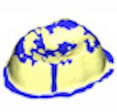
At RWTH Aachen, 3D ultrasonic scan strategies are being optimized and evaluated. Based on this work, an ultrasonic microscanner prototype is being developed, along with control modules. In the oral cavity, a miniaturized ultrasonic scanner can capture and display margins regardless of presence of blood, saliva, or gingiva, according to Radermacher.
Ultrasound can penetrate periodontal tissue, saliva, and blood, helping improve cast precision for the CAD/CAM manufacturing process. An ultrasound-based method is therefore a noninvasive method of intraoral digital impressions with no adverse effect on the periodontium, he and his team explain on the project website.
Methods for sound coupling are being evaluated against optical reference scans provided by BEGO Medical, one of the project partners. BEGO also produces corresponding crowns and bridge framework for precision control and integrates the scanner into the CAD/CAM process chain via an open interface necessary for OEM production.
The Aachen University Department of Prosthodontics is also involved in the project, providing tooth samples, casts, and other material, and evaluating tooth preparation and the precision and location of implants.
SurgiTAIX, a developer of technologies for minimally invasive surgery, is providing, evaluating, and integrating high-frequency ultrasonic transmitter and receiver modules and scanner stabilizing positioning systems for the project. A splint fixing the scan unit to the dentition helps avoid in-motion artifacts and ensures stability.
Preliminary results confirm the precision of axial scanning to match the design specifications, according to Radermacher and his team.
 A schematic of the intraoral ultrasonic microscanner being developed at Aachen University. Image courtesy of RWTH Aachen.


