Imaging assisted in the diagnosis and removal of a dental drill head from the airway of a 76-year-old patient who aspirated the object during a procedure, according to a case report recently published in Radiology Case Reports.
An x-ray and computed tomography (CT) scan aided in visualizing the approximately 2-cm dental drill head within the patient's right airway. Once the foreign object was retrieved, the patient recovered without incident, the authors wrote.
"This case underscores the importance of thorough diagnostic evaluation and the use of precise imaging modalities in patients with suspected foreign body aspiration, even in the absence of symptoms," wrote the authors, led by Dr. Marco De Chiara of the University of Campania in Naples, Italy (Radiol Case Rep, March 8, 2025, Vol. 20:5, pp. 2408-2411).
A 76-year-old patient with a foreign body ingestion sensation
In March 2024, the patient went to an emergency department with concerns about ingesting something during a dental procedure. The patient had no symptoms, including respiratory distress, abdominal pain, or trouble swallowing. The patient's vital signs were stable, a clinical exam was unremarkable, and an electrocardiogram revealed no signs of heart abnormalities, according to the report.
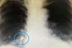
First, a chest x-ray was completed, which showed a 2-cm metallic foreign body within the patient's right airway. The object was consistent with a dental drill head that had been aspirated, the authors wrote.
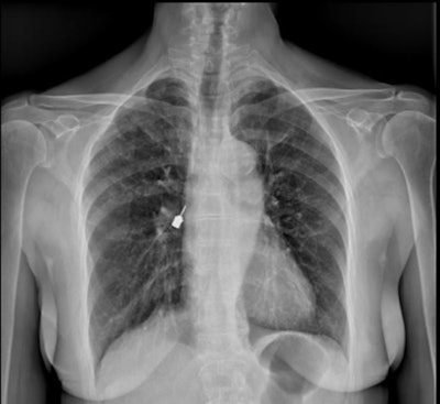 Figure 1: Posteroanterior chest x-ray. A metallic foreign body (the drill head) is visible near the right hilum, supposedly located in the right airways. Images and captions courtesy of De Chiara et al. Licensed under CC BY-NC-ND 4.0.
Then a CT scan was completed to get a precise location of the drill head. The scan confirmed that the drill head was in the intermediate bronchus and extended into the inferior bronchus on the right side.
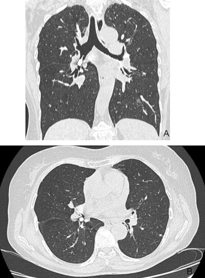 Figure 2: Thoracic CT scan showing the drill head located in the right intermediate and right lower bronchi. (A) coronal view (B) axial view.
The patient underwent an urgent bronchoscopy for retrieval. The drill head was removed without significant mucosal trauma, they wrote.
The patient developed a mild postprocedural cough and hoarseness for about two days but otherwise recovered without complications.
Early diagnosis and intervention are key
The aspiration of dental instruments, including endodontic files, implant screws, and drill heads, is rare, with its incidence estimated to range from 0.004% to 0.007% during dental procedures. Patients undergoing procedures in a reclined position and those with preexisting respiratory conditions may be at greater risk of aspiration, the authors wrote.
Early recognition and accurate diagnosis are important to prevent airway obstruction, pneumonia, atelectasis, or chronic inflammation leading to bronchiectasis.
Therefore, dental professionals need to mitigate these risks and better patient safety, they wrote.
"This case provides an educational opportunity for dental practitioners and radiologists, emphasizing the importance of recognizing and managing foreign body aspiration promptly,” De Chiara and colleagues wrote.

