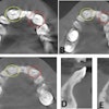
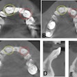
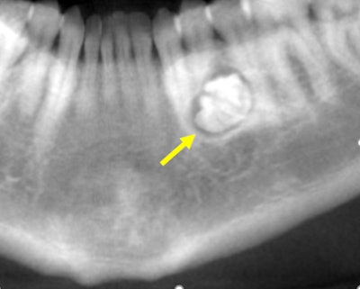
 Figure 1: Reformatted CBCT panoramic film.
DrBicuspid publishes a new case study weekly. To test your dental expertise, first, please log in. If you don't have a login, create one. Each case comprises a history, quiz, and discussion section. An answer key is provided at the end of the case study.
Log in to view the full article
 Figure 1: Reformatted CBCT panoramic film.
DrBicuspid publishes a new case study weekly. To test your dental expertise, first, please log in. If you don't have a login, create one. Each case comprises a history, quiz, and discussion section. An answer key is provided at the end of the case study.
History: The patient is a 32-year-old woman who was referred to the oral surgeon by her dentist because of a lesion on the left mandible, which was noted in a panoramic film. The patient was asymptomatic and had no paresthesia or swelling.
Her past medical history was significant for seasonal allergies. The intraoral and extraoral exams were within normal limits.
The oral surgeon ordered a cone-beam computed tomography (CBCT) scan and requested an interpretation report by an oral radiologist. Images are provided below in the following order:
- CBCT cropped panoramic film
- CBCT coronal projections (anterior to posterior)
- 3D reconstruction of the left mandible
 Figure 1: Reformatted CBCT panoramic film.
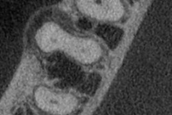
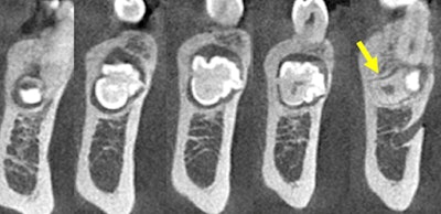 Figure 2: CBCT coronal projections (anterior to posterior).
The oral radiologist provided a radiographic interpretation report to the dentist. A paragraph of the report is provided below:
A hyperdense lesion (radiopaque) surrounded by a hypodense ring with very well-defined
borders. A distal extension of the lesion takes the form of a primitive root. The lesion is in the middle of the mandible. Tooth #21 is slightly mesially displaced. Initial resorption is observed at the distal third aspect of the root of tooth #21. The lesion is displacing apically the mental foramen.
1. What is the LEAST likely differential diagnosis?
A. Idiopathic bone sclerosis
B. Simple bone cyst
C. Cemento-osseous dysplasia
D. Cementoblastoma
E. Odontoma
2. The radiologist included three differential diagnoses in the report, including an odontoma. What is the most likely differential diagnosis suggested by the radiologist?
A. Complex odontoma
B. Compound odontoma
C. Fibro-odontoma
D. None of the above
3. What is the next best step?
A. Incisional biopsy
B. Excisional biopsy
C. Prescription of antibiotics
D. Surgical excision of the lesion
Discussion
Odontomas typically present in the first and second decades of life, and they are accepted as developmental anomalies (hamartomas) rather than true neoplasms. The etiology of odontomas remains unknown. Several theories have been proposed, and various causes -- including trauma, infection, family history, and genetic mutation -- have been postulated. Some investigators have suggested that the ameloblastic fibroma and ameloblastic fibro-odontoma both developmentally and histomorphologically represent the early stages of the formation of odontomas.
Most odontomas are asymptomatic, and radiographic findings are by and large diagnostic. Usually, the compound odontoma appears as a collection of toothlike structures surrounded by a narrow radiolucent zone. Hence, it can seldom be confused with any other entity such as supernumerary teeth. Occasionally, it may become large and produce expansion of bone with consequent facial asymmetry.
Odontomas must be surgically removed to prevent cyst formation and possible conversion to odontoameloblastoma. Ameloblastic odontoma and ameloblastic fibro-odontoma bear a great resemblance to the common odontoma, particularly on the radiograph; thus, it is suggested that all odontomas should be sent to an oral pathologist for microscopic examination and definitive diagnosis. In most odontomas, pathologic alterations are observed in the neighboring teeth, such as devitalization, malformation, aplasia, malposition, and remaining embedded.
Most odontomas are discovered accidentally, thus further supporting the use of radiography as an indispensable tool in routine dental clinical examination.
Reference
Neville BW, Damm DD, Allen CM, Bouquot JE. Oral and Maxillofacial Pathology. 3rd ed. St. Louis, MO: Saunders/Elsevier; 2009.
Answer key
1. B: simple bone cyst
2. B: compound odontoma
3. D: surgical excision of the lesion

