Imaging, specifically cone-beam computed tomography (CBCT), may improve dental implant planning and lower the risk of complications by identifying pathologies before procedures. The study was recently published in BMC Oral Health.
Also, CBCT may be key in detecting sinus abnormalities and aid in effective planning for sinus augmentation in implant patients, the authors wrote.
“This study highlights the high prevalence of sinus pathologies in dental implant candidates and their significant association with odontogenic factors,” wrote author Sercan Küçükkurt, PhD, of Istanbul Aydın University (BMC Oral Health, May 23, 2025, Vol. 25, 776).
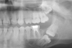
The study analyzed 1,000 CBCT scans from 500 men and 500 women, examining a total of 2,000 maxillary sinuses. Both sinuses were reviewed for each patient, even if only one side required an implant. Panoramic radiographs provided an initial overview of the posterior maxilla followed by detailed coronal and sagittal CBCT reconstructions for thorough sinus evaluation, Küçükkurt wrote.
Sinus pathologies were categorized based on demographic differences and dental factors of periapical lesions with or without root canal treatment (RCT), RCT teeth without lesions, and edentulism. Researchers also noted whether these conditions were unilateral or bilateral and used statistical analysis to explore their association with sinus abnormalities.
Sinus pathologies were found in 39.5% of sinuses and affected 54.8% of patients. Mucosal thickening was the most common condition (61%) followed by cysts or polyps (27.6%) and opacifications (11.4%). Men had a significantly higher rate of cysts and polyps (p = 0.020), while mucosal thickening did not vary by gender, Küçükkurt wrote.
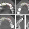
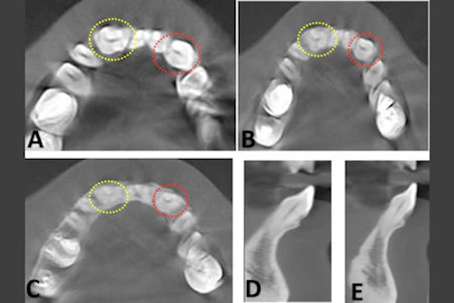
 Maxillary sinus conditions identified via CBCT. (A) Clear sinus, (B) mucosal thickening, (C) cyst/polyp, (D) opacification (≥ 50% of sinus volume). Images and captions courtesy of Küçükkurt. Licensed under CC BY-NC-ND 4.0.
Furthermore, odontogenic factors were present in 65.2% of affected sinuses, especially in mucosal thickening cases, with periapical lesions, both untreated and treated with RCT, strongly linked to pathology (p < 0.0001 and p = 0.013), unlike RCT teeth without lesions (p = 0.411). Complete sinus opacification occurred in 5% of cases, and patients ages 41 to 60 had a significantly higher rate of bilateral sinus involvement (p < 0.0001). Mucosal thickening was more often bilateral, while cysts were typically unilateral (p = 0.003).
However, the study had limitations. The absence of follow-up data prevented evaluation of how dental treatments may impact sinus health over time, Küçükkurt added.
“These findings underscore the indispensable role of CBCT in identifying sinus abnormalities, facilitating accurate diagnosis, and optimizing sinus augmentation and implant planning,” he concluded.