On-screen evaluation of color 3D models may show strong diagnostic agreement with traditional visual examinations for detecting caries in children, according to a study published in JMIR Public Health and Surveillance.
However, the extent to which on-screen assessment of intraoral scanner (IOS)-generated 3D models can replace visual examinations may depend on the clinical context, the authors wrote.
“On-screen assessment of 3D models in color supplemented with fluorescence had a high level of agreement with visual examination for detecting primary coronal carious lesions on smooth and occlusal surface types at the moderate and extensive disease thresholds,” wrote the authors, led by Bree Jones, PhD, MPH, of the University of Melbourne Dental School in Australia (JMIR Public Health Surveill, December 8, 2025, Vol. 11, e78023).
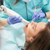
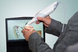
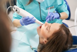
A total of 216 children, with an average age of approximately 6, completed a clinical trial that included visual examinations using the International Caries Detection and Assessment System (ICDAS) and intraoral scanning with the Trios 4 system. Visual exams were conducted, and all tooth surfaces were assessed. Immediately after the visual exam, all teeth were scanned with the Trios 4 IOS, they wrote.
Eight months later, dental practitioners independently evaluated the color 3D models and then models supplemented with fluorescence, using a validated, modified ICDAS index, with reassessments performed after four weeks. Caries were recorded at the tooth-surface level, with outcomes categorized into initial, moderate, and extensive disease thresholds, and assessment times were documented.
The mean time required to complete an on-screen assessment of each color 3D model with fluorescence was 3.5 minutes. Caries detection using color 3D models showed similar odds to visual examination across all thresholds, including initial (odds ratio [OR], 1.1; 95% confidence interval [CI], 1 to 1.3), moderate (OR, 0.9; 95% CI, 0.7 to 1.1), and extensive disease (OR, 1; 95% CI, 0.7 to 1.3), they wrote.
When fluorescence was added, caries detection was 30% more likely at the initial threshold compared with visual examination (OR, 1.3; 95% CI, 1.1 to 1.5) while remaining similar at moderate and extensive thresholds. Further analyses demonstrated strong agreement between methods at moderate and extensive levels. Overall agreement was high (intraclass correlation coefficients [ICC], 0.9; 95% CI, 0.9 to 1), with intra- and interexaminer reliability ranging from good to excellent (ICC, 0.8 to 1).
However, the study was limited by the incomplete recording of examination times for all participants. Future research should consistently document this information to better assess its clinical relevance, the authors added.