Practitioners should use 3D imaging prior to removing a lower third molar in patients with a high risk of inferior alveolar nerve (IAN) injury, according to a new study in Oral Surgery, Oral Medicine, Oral Pathology, Oral Radiology, and Endodontics (October 15, 2010).
In a retrospective case series study, researchers from University Hospital of Zurich and Harvard Medical School chose a study population composed of patients presenting with an impacted lower third molar with projection of the tooth over the full width of the IAN in a panoramic radiograph. They considered the spatial relationship to the IAN, type of angulation, root configuration, and maturation.
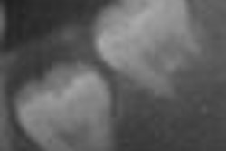
A total of 707 wisdom teeth in 472 patients were evaluated using 3D imaging. A close relationship to the IAN was seen in 69.7% of the teeth, and the diameter of the mandibular canal was reduced in 45.1%. In 52.8%, the IAN was vestibular and in 37.3% lingual to the roots; there were 9.9% with an inter- or intraroot course. Most teeth had 1 or 2 roots (86.7%), but 13.3% had 3 or more roots. Mesial angulation was the main type (40.2%), followed by vertical (29%), horizontal (13.9%), distal (10.2%), and transverse (6.8%) positions.
Based on the range of variations in the course of the nerve and the number of roots, the authors recommend 3D imaging before surgical removal of a lower third molar that shows signs of a close relationship to the IAN.
Copyright © 2010 DrBicuspid.com