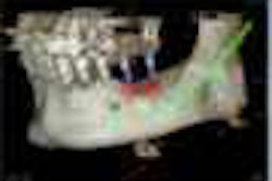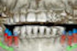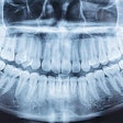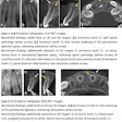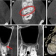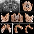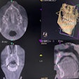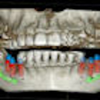
Cone-beam CT (CBCT) appears to be as accurate as 2D panoramic radiography for predicting whether the inferior alveolar nerve (IAN) will be exposed during mandibular third-molar extraction in adults.
A team from the University of Toronto presented these results from a systematic literature review February 14 at the Faculty of Dentistry's 2012 Research Day.
Third-year dentistry student Adam Ohayon and five others searched three electronic databases and retrieved 79 articles. They closely examined the details of nine of the articles, focusing on the comparative accuracy of CBCT and panoramic radiography for predicting IAN exposure among adults with impacted mandibular third molars. They then compared the two articles that remained (Oral Surgery, Oral Medicine, Oral Pathology, Oral Radiology and Endodontology, February 2007, Vol. 103:2, pp. 253-259, and International Journal of Oral & Maxillofacial Surgery, September 2009, Vol. 38:9, pp. 964-971).
'Grade A evidence'
The first article was by members of the Department of Oral and Maxillofacial Radiology at Tokyo Medical and Dental University. The researchers prospectively studied the imaging of 42 impacted mandibular third molars in 135 patients. They used J. Morita panoramic radiography (Super Veraviewepocs) and CBCT (3DX) systems and conducted the panoramic radiographic imaging two weeks before CBCT imaging.
Two of the team members -- whom Ohayon and his group found to be "highly qualified [image] evaluators" and who had not removed the teeth -- were presented with CBCT images in a randomized order so they could not refer to the panoramic features.
There was an 80% accuracy rate with CBCT compared to the gold standard of direct intra-operative visualization of IAN exposure, including 93% sensitivity and 77% specificity. There also was "excellent" interobserver agreement.
PAN's overall accuracy was 64% (p < 0.05 versus CBCT), with 70% sensitivity and 63% specificity, and there was "fair to excellent" interobserver agreement.
Ohayon's team concluded the study provided "Grade A evidence" for the relative accuracy of CBCT. The study's main weakness was possible selection bias due to exclusion of teeth for which there was limited direct intraoperative visualization, they noted.
Selection bias
Researchers from the Department of Oral and Maxillofacial Surgery at The Radboud University Nijmegen Medical Centre conducted the other prospective, blinded study.
The investigators -- who were "experienced" and "qualified" image evaluators -- analyzed the imaging results from 53 impacted mandibular third molars in 40 patients. CBCT (i-CAT 3-D Imaging System, Imaging Sciences International) had a 55% accuracy compared to direct intra-operative visualization. CBCT's sensitivity was 96%; its specificity was 23%; and there was excellent inter-observer agreement.
The overall accuracy of their panoramic radiography system (Cranex Tome, Soredex) was 45%, with 100% sensitivity and 3% specificity. There was poor interobserver agreement.
The Toronto team determined that this study provides "Grade C evidence" for CBCT's superiority because the investigators did not provide a p-value for the CBCT-PAN comparison. Also, as Ohayon pointed out, there appears to be selection bias in the study.
"They only included teeth for comparison that had obvious radiographic indicators on the panoramic radiograph that there would likely be an IAN exposure during extraction," he said. "If you're going to choose teeth with such obvious indicators, then what extra significant diagnostic benefit will CBCT truly provide?"
From their analysis, Ohayon and his colleagues concluded that while CBCT is as good as panoramic radiography in diagnosis of IAN exposure, its 3D imaging capability "may provide for better overall preoperative assessment, informed consent, and indication of the surgical technique, as the IAN can be assessed in a buccal and lingual direction, as well."
