Orthodontic patients pose some unique challenges when it comes to cone-beam CT (CBCT) imaging, and dental practitioners should exercise caution to ensure they don't expose these patients to more radiation than is diagnostically necessary, according to the president of the American Academy of Oral and Maxillofacial Radiology (AAOMR).
Orthodontists were some of the earliest adopters of cone-beam CT in dentistry, according to Allan Farman, B.D.S., M.B.A., Ph.D., D.Sc., who in addition to being the current AAOMR president is a professor of radiology and imaging science at the University of Louisville in Kentucky. This is not surprising given the technology's ability to more precisely identify anatomical and morphological details than 2D imaging and to do so at a much lower dose than medical CT.
But Dr. Farman believes there is a dichotomy in the orthodontic community regarding the value of 3D imaging for orthodontic planning and the number and type of images needed to optimize outcomes.
"On the one hand, there were people (mainly on the West Coast), the earliest adopters, who decided before the dosimetry studies came out that everyone should get a 3D image," he said. "And those individuals are going to be difficult to convince that they may have jumped the gun. On the other extreme are those people, including orthodontists, who don't like taking any radiographs at all on this young population. And then there are those in the middle who say that sometimes 3D is warranted, but not in 100% of the cases."
“We are dealing with different issues with regard to the ill effects of radiation on this younger age group.”
— Allan Farman, president, American
Academy of Oral and Maxillofacial
Radiology
The key, according to Dr. Farman, is for dental professionals to minimize the dose and maximize the quality of information available for specific tasks. This is especially important in orthodontics, he added, where the majority of patients are younger and thus more susceptible to radiation damage over their lifetime, and the entire head is often imaged four times: before, during, and after treatment, and then again to see if treatment is being retained.
"A child or young teen is three times more susceptible to radiation volume than a young adult, who is three times as susceptible as someone in their 50s and 60s," he said. "So we are dealing with different issues with regard to the ill effects of radiation on this younger age group."
Stuart C. White, D.D.S., Ph.D., a professor at the University of California, Los Angeles School of Dentistry and chair of the school's oral radiology section, agreed.
"We should be exposing patients only when there is a reasonable likelihood that the examination will yield information that will change the patient's diagnosis or treatment plan," Dr. White said. "Many orthodontic patients can seemingly be treated perfectly well without cone-beam CT examinations (and have been for many decades), although some patients certainly benefit -- hence the need for guidelines to identify those patients likely to benefit and those likely not to benefit."
Collimation and dose differences
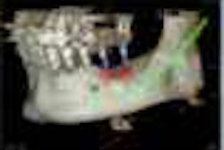
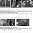
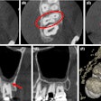
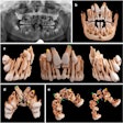
For example, Dr. Farman noted, "It is not unusual for the maxillary canine in an orthodontic patient to be impacted, and those impacted canines probably do need to be viewed in 3D to get a better idea of the exact location and determine whether it is possible to move them into place with orthodontic force."
In fact, the authors of a recent Seminars in Orthodontics article concluded that "the use of CBCT in orthodontics greatly enhances our understanding of impacted canines and offers unique, comprehensive information for individual situations. Compared with conventional imaging approaches, the fidelity of this information is unsurpassed" (September 2010, Vol. 16:3, pp. 199-204).
But this doesn't mean you have to do a full-head cone-beam CT scan when all you really want to look at is the canines, Dr. Farman noted. "You could collimate the beam down to just a few centimeters' heights," he said. "Six centimeters or less would be quite adequate."
The problem is that not every commercially available cone-beam CT system offers collimation, he noted.
"What sort of cone-beam CT system should an orthodontist buy? They shouldn't buy one that won't collimate because they should only be imaging those tissues that need to be examined," Dr. Farman said. "You should not expose an area greater than what you need to expose, and there is no reason to have a beam larger than the area of interest. Some cone-beam CT systems allow collimation and some don't, and I am fairly negative about those that don't."
Dosages vary considerably from system to system as well, he added, depending on how old the product is and whether it is full or partial field-of-view.
"There is a tremendous range in the dose supplied by different cone-beam CT systems," Dr. Farman said. "The Hitachi MercuRay and early versions of Iluma and ProMax systems were relatively high dose compared to other cone-beam CT systems. But this is changing, and Hitachi withdrew from the cone-beam CT maxillofacial market altogether. But these older machines are still out there, and the FDA has not outlawed them."
Some newer systems, such as the iCAT, the Galileos, and the NewTom, are considered low dose, although not all of them (such as the NewTom) can be collimated, he added.
The bottom line is that practitioners need to limit as much as possible the amount of radiation a patient is exposed to while maximizing the diagnostic information.
"And they need to be aware that, even if they are told at a meeting that a cone-beam CT scan is equal in radiation dose to a single airport x-ray or ultrasound scan, it is three orders of magnitude wrong," Dr. Farman emphasized. "And whether you are taking a periapical film or a cephalograph or a cone-beam CT scan, you need to use professional judgment on whether the image is warranted."
Legal liabilities
This same advice applies to the potential legal liabilities associated with image interpretation, Dr. Farman added. A recent article by the legal counsel for the American Association of Orthodontists (AAO) noted that "a plethora of issues unrelated to dental and orthodontic matters can be revealed by cone-beam films. Orthodontists may not be trained to interpret all of the cone-beam data" (Bulletin, August 2010, Vol. 28:5, pp. 24-25).
In particular, the article noted, "if AAO members elect to interpret the cone-beam information on their own, they will have accepted a greater duty to the patient than that to which they would otherwise be obligated. In other words, if the cone-beam data reflects any condition or issue other than a dental or orthodontics matter, the orthodontist will have assumed the duty to accurately identify it."
The article goes on to recommend that orthodontists utilize the services of someone trained to properly interpret cone-beam films, such as an oral and maxillofacial radiologist.
"The question posed by the AAO legal counsel was whether or not orthodontists should spend time reviewing these images or send them out for review," Dr. Farman said. If the image is a full field-of-view, he recommends that those views be sent out for a second opinion by someone better qualified to interpret them. "So that for the sake of the patient, and the practitioner's ethical duty and possible legal issues, one should go back and make sure that the volume of information is read more fully so the patient can benefit maximally from the dose."
The AAOMR is currently working with the AAO to develop imaging use guidelines for orthodontics, he noted.
Copyright © 2010 DrBicuspid.com
