Cone-beam CT (CBCT) gets the royal treatment in a series of articles published in the January 2010 Journal of the California Dental Association (Vol. 38:1). Having been used in oral and maxillofacial imaging for more than a decade, cone-beam CT is gaining ground in clinical use, with new applications ranging from diagnosing dental disease to evaluating the temporomandibular joint (TMJ) and enhancing maxillofacial pathology.
"Although CBCT technology was originally introduced as state-of-the-art imaging, it is entering the mainstream of everyday dentistry, enriching the diagnostic armamentarium of dental practitioners," wrote Sotirios Tetradis, D.D.S., Ph.D., and Stuart C. White, D.D.S., Ph.D., in their introduction.
Disease diagnosis
In "Cone Beam Computed Tomography in the Diagnosis of Dental Disease," Dr. Tetradis and co-authors Paul Anstey, D.D.S., and Steven Graff-Radford, D.D.S., noted that while conventional radiographs provide important diagnostic information for caries detection, periodontal evaluation, endodontic applications, root resorption, and trauma to the teeth, they represent 2D images of 3D objects with significant structure superimposition and unpredictable magnification.
Cone-beam CT, on the other hand, allows true 3D visualization of the dentoalveolar structures, avoiding major limitations of conventional radiographs and offering advantages in disease detection for selected patients, they wrote.
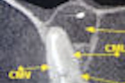
In "Cone Beam Computed Tomography Imaging in the evaluation of the Temporomandibular Joint," the authors noted that accurate evaluation of the TMJ using conventional x-rays has historically been difficult due to the superimposition of other structures. Cone-beam CT, however, "provides precise imaging of TMJ anatomy without superimposition and distortion."
"CBCT has become the imaging of choice for presurgical evaluation in surgery, for dental implants, and is replacing older imaging modalities for evaluation of TMJ disease," they wrote. "Although CBCT does not image soft-tissue, such as disc position relative to the condyle and fossa, appropriate clinical evaluation can usually determine if a disc displacement is a factor that needs to be treated."
Legal liabilities
Still, the technology is not without its controversies. In "Legal Considerations in the Use of Cone Beam Computed Tomography Imaging," Edwin Zinman, D.D.S., J.D.; Dr. Tetradis; and Dr. White address questions regarding the legal responsibilities associated with the use of cone-beam CT, including cone-beam CT necessity, recognition of pathosis in the scan's entire volume, adequate training, and informed consent and/or refusal.
"In conjunction with the advantages and opportunities from the application of new technologies in patient care, responsibilities and obligations for proper use of such technologies also emerge," they wrote.
They pose a number of questions relating to the legal liabilities of dental professionals who order and read cone-beam CT exams, such as "Is a dentist legally obligated to recognize or diagnose all disease evident in a cone-beam CT examination if it is not in the field of interest for which the image was ordered?" and "Can a dentist be liable for not ordering a cone-beam CT or other volumetric examination?"
In the long run, the authors recommend erring on the side of caution.
"Dentists should use CBCT as an advanced diagnostic tool to aid diagnosis and treatment planning when indicated," they wrote. "The dentist should obtain hands-on learning to appreciate the diagnostic information contained in the CBCT image or refer the patient to an expert. Dentists have a legal and ethical obligation to provide and protect the patient's best interest as their primary goal in patient care."
Copyright © 2010 DrBicuspid.com