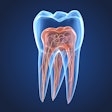
Effective root canal therapy depends on cleaning out all the infected canals. But these canals can be hard to find, let alone negotiate. Often the only choice is to cut away tooth structure in search of lumens narrower than a human hair.
So a new report that cone-beam CT is highly successful in finding root canals could prove important.
"An attempt to locate them can result in increased chair time and perforate the tooth or root," said Nadia Chugal, D.D.S., M.S., M.P.H., a University of California, Los Angeles professor of endodontics. "Searching for them, you can remove a lot of tooth structure you don't want to remove. So it helps to have good imaging. And conventional radiography is not sufficient."
That's why Dr. Chugal and her colleagues became interested in cone-beam CT. It moves on multiple planes as it creates a 3D image, potentially catching something that would not appear on the single plane view offered by conventional x-rays. "Could it tell us whether there are additional canals, how many, and where they are?" Dr. Chugal wondered.
To test the idea out, the team used microcomputed tomography (micro-CT) as the gold standard. Micro-CT is not possible to use inside a living mouth, but Dr. Chugal's team used it on extracted teeth to verify the accuracy of cone-beam CT versus conventional radiographs.
For their study, presented at the recent International Association for Dental Research (IADR) meeting in Miami, they analyzed 43 teeth using a 3DX Accuitomo (J. Morita) and periapical F-speed film radiographs, then confirmed the findings with micro-CT.
Five evaluators participated: two radiologists and three endodontists.
The researchers found that the cone-beam CT findings agreed with those of micro-CT 98.6% of the time, while the periapical radiograph only agreed 69.0% of the time. The difference was significant (p < 0.05).
They also found a significant difference in the level of agreement between observers using cone-beam CT versus periapical radiographs (p ≤ 0.007). And they found that the detection of distance of the canal lumen from the cementoenamel junction was significantly shorter in cone-beam CT scans compared to periapical radiograms (p < 0.05).
"The 3DX Accuitomo images proved to be superior in its ability to localize canal lumina compared to conventional periapical radiographs," Dr. Chugal and her colleagues concluded.
The research supports other recently published work, including a study by researchers at the universities of Joinville and Positivo who compared cone-beam CT (i-CAT, Imaging Sciences International) to ex vivo and clinical methods for finding root canals. "CBCT can be used as a good method for initial identification of maxillary first molar internal morphology," the researchers concluded (Journal of Endodontics, March 2009, Vol. 35:3, pp. 337-342).
The lead author of the Brazilian study, Flares Baratto Filho, Ph.D., stated in an e-mail that Dr. Chugal's study "has important results and it can be useful in endodontics."
But he warned that cone-beam CT won't solve all the problems of calcified canals. Many can't be negotiated even after they are identified, he noted. "The experience of operator is very important in this case," he said.
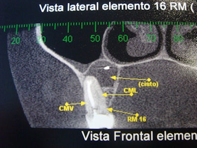 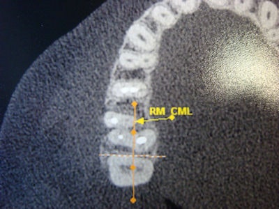 |
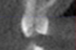
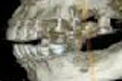
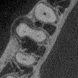
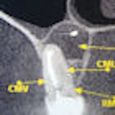
| Left, a cone-beam CT lateral view showing a fractured root canal in a first maxillary premolar. Right, a cone-beam CT occlusal view showing root canals. Images courtesy of Flares Baratto Filho, Ph.D. |
Copyright © 2009 DrBicuspid.com


