A common treatment in the modern dental practice is replacing a single posterior tooth with a dental implant. I am able to place an implant with confidence and precision in less time than it takes to excavate and fill a two-surface resin restoration or a single rooted endodontic procedure.
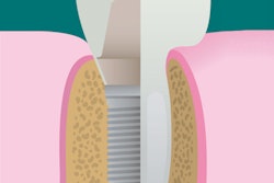
This happens routinely as I perform computer-assisted guided implantology. Executing a flapless or miniflap-guided implant surgery benefits my patients in a multitude of ways. I begin with advanced treatment planning with Cerec Galileos (Sirona) integration, which prepares me for prosthetically driven implant surgery. I use this protocol for all my cases, and it yields many advantages over conventional implant surgery. The goal is to perform a safe and optimally positioned installation of the implant, which becomes easily restorable.
The benefits of guided implantology include the following:
 Anthony Ramirez, DDS.
- Decreased surgical time
- Faster postoperative healing
- More precise and accurate implant positioning
- Less surgical trauma through a tissue punch access
- Uncomplicated post-op
- Less chance of penetrating vital anatomical structures because of 3D planning
- Working with a fixture that is easy to restore
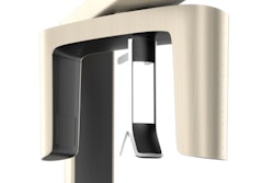
A 2012 position statement by the American Academy of Oral and Maxillofacial Radiology recommends that cross-sectional imaging be used for the assessment of all implant sites and that cone-beam CT (CBCT) is the imaging method of choice for gaining this information.
Approaching a case with the precise planning via CBCT imaging removes a big variable -- the unknown -- and increases the opportunity for success.
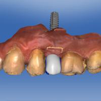
Selecting a patient who has adequate bone volume and ample keratinized tissue for the procedure is important. The ideal position for the implant relative to the subsequent prosthetic tooth replacement can be planned virtually. If necessary, perform bone and connective tissue grafts to produce the bone volume and provide the best tissue before implant installation. The position of the implant is calculated as a direct result of a Cerec crown proposal from an Omnicam optical scan of the dentition that is merged with the radiographic scan data from the Galileos CBCT.
The following two cases illustrate treatment with two types of guides and how the streamlined digital workflow creates a methodical approach in my hands that is the foundation for my clinical success. Case No. 1 used a SICAT surgical guide that was fabricated from the data I uploaded and transferred through the global portal to the SICAT laboratory in Germany. Case No. 2 was treated with an in-office Cerec Guide 2 that is designed in Cerec and milled in my MC XL chairside milling unit (Sirona).
Both guides are accurate at producing osteotomies precisely planned from the implant software. The cost of a surgical guide has never been a deterrent to case acceptance and is actually welcomed by my patients when I explain all the benefits and why I am using it.
Why go through the workflow as I have outlined and not treat these cases as simple freehanded implant surgeries? Because I have seen many implants that are off-angled, difficult to restore, and poorly positioned, in close proximity to adjacent teeth when placed in that manner. Cross-sectional imaging using CBCT can be eye opening when reviewing the results of previously placed implants.
Even the most experienced implant surgeons can't duplicate the precision of properly planned and installed implants when using surgical guides. I want my patients to have the benefit of accurately positioned, well-integrated implants that can be long-lasting in function, and that's why I choose to work with surgical guides in all of my implant cases. CBCT imaging is the basis for success and helps me avoid some of the pitfalls of conventional implant surgery when performed with limited 2D radiographic imaging. Knowing what you are up against before commencing with surgery will help you avoid penetrating vital anatomical structures, such as atypical anatomy, boney defects, sinuses, nerves, adjacent teeth, and others.
Case 1
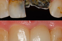
A healthy 45-year-old patient was treatment planned for Invisalign clear-aligner therapy to allow for the replacement of a missing tooth No. 13 and improve the overall stability of his occlusion. The resultant space created was adequate for a 3.3 x 14-mm Straumann SLActive NC bone level implant (Straumann) and would challenge even the most experienced surgeon.
Planning and working with a SICAT Optiguide (Sirona) provided certainty and made this case manageable. The images speak for themselves, and I expect this implant to be long-lasting, functioning well under an aesthetically pleasing screw-retained hybrid abutment crown.
Case 2
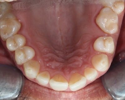
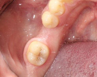
Treatment was planned for a healthy 25-year-old patient for multiple implants to replace two hopeless maxillary first molars and a missing mandibular left first molar. The implant site at No. 19 was slightly deficient at the crest, and I preplanned to use a miniflap approach to install a 4.8 x 12-mm Straumann SLActive RC bone level implant and place a concomitant buccal contour bone graft to add crestal bone at the time of implant placement. The procedure was completed via an in-office Cerec Guide 2, which was digitally designed and milled from a block of polymethyl methacrylate (PMMA) material. The osteotomy was prepared with the Cerec Guide keys for the Straumann guided system.
Positive impact
Clarity of vision, minimally invasive, guided implantology is sure to positively affect any implant practice -- which, in short, justifies and furthers the ample advantages of practicing in the third dimension.
Anthony Ramirez practices in Brooklyn, NY. He can be reached at [email protected] or via his website.
The comments and observations expressed herein do not necessarily reflect the opinions of DrBicuspid.com, nor should they be construed as an endorsement or admonishment of any particular idea, vendor, or organization.
