The evaluation of buccal bone can be difficult as panoramic or periapical images may underestimate the amount of bone available. Cone-beam CT (CBCT) units can be useful for making an accurate diagnosis, but is any particular system on the market more accurate?
Researchers tested six CBCT systems in a blinded in vitro study. While all six were highly accurate when detecting buccal bone loss, one unit showed slightly higher accuracy, they found (Australian Dental Journal, March 6, 2019).
"All six CBCT units provided high diagnostic accuracy in the evaluation of [buccal bone]," wrote the authors, led by Dr. Luciana Loyola Dantas of the department of prosthetics and integrated clinics at the Federal University of Bahia School of Dentistry in Brazil.
Accuracy
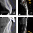
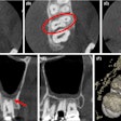
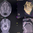
When attempting to visualize the amount of buccal and lingual bone present in a patient's mouth, both periapical and panoramic images have limitations, the authors wrote. Cone-beam CT allows practitioners to assess buccal bone and any anomalies, such as dehiscence and fenestration, in ways that other imaging modalities do not allow.
“All six CBCT units provided high diagnostic accuracy.”
While CBCT's applicability in imaging buccal bone has been studied before, this is the first study that compares multiple devices in their ability to detect the presence of buccal bone, according to the authors. Until the present, no studies have reported on the diagnostic accuracy of different CBCT systems in assessing buccal bone in anterior teeth, they noted.
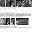
The researchers wanted to judge the accuracy of six CBCT systems in assessing how much buccal bone is present in anterior teeth. They covered both jaws of a human skull in wax and submerged it in water for 24 hours before image acquisition.
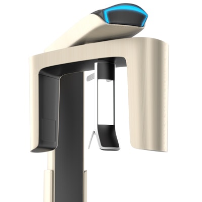
The researchers took images of the skull with six different CBCT systems:
- 3D Accuitomo 170 (J. Morita)
- CS 9000 3D (Carestream Dental)
- CS 9300 (Carestream Dental)
- Eagle 3D (Dabi Atlante)
- i-CAT Classic (i-CAT)
- Orthophos XG 3D (Dentsply Sirona)
They based exposure and acquisition protocols on the manufacturers' guidelines, with voxel size adjusted to be as close as possible to 0.2 mm. They then randomly evaluated cross-sectional images when the buccal bone was assessed. All images were blindly evaluated by two oral radiologists who did not know about the CBCT systems or the diagnosis.
All of devices demonstrated high accuracy in detecting the loss of buccal bone, but the images from the CS 9300 showed slight superiority in accuracy (see table below), the researchers found. However, the differences were not statistically significant, they noted.
| Comparison of accuracy of 6 CBCT systems by observer | ||||||
| Orthophos | CS 9000 3D | i-CAT | Eagle 3D | 3D Accuitomo 170 | CS 9300 | |
| Observer 1 | 88.9% | 88.9% | 91.7% | 91.7% | 91.7% | 94.4% |
| Observer 2 | 75.0% | 86.1% | 86.1% | 88.9 | 88.9% | 91.7% |
Radiation dosage
Among the limitations mentioned by the authors was that this was in vitro study. Issues such as image artifacts, trauma, and gingival recession were not present in this evaluation.
They concluded that all six CBCT systems tested provided high diagnostic performances for evaluating buccal bone in anterior teeth. However, the radiation dose to which the patient is exposed is an important consideration, they noted.
"When the clinical usefulness of an imaging modality is equivalent, choice of appropriate imaging should be directed toward the modality that delivers the least patient radiation dose for a specific diagnostic task," the authors wrote.
