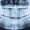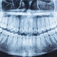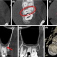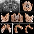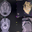
Is an image from a panoramic radiograph sufficient, or is a cone-beam CT image needed for an accurate diagnosis? Researchers asked residents in oral surgery and orthodontics this and other questions to measure imaging accuracy and more.
Both sets of residents reported that they considered the CBCT images to be more accurate. The study was published in the International Journal of Implant Dentistry (December 2018, Vol. 4:37).
"Overall diagnostic accuracy is decent with [panoramic radiographs] and can be improved with CBCT," wrote the authors, led by Josipa Radic of the Clinic of Cranio-Maxillofacial and Oral Surgery at the University of Zurich Centre of Dental Medicine in Switzerland.
Image accuracy
Although there is considerable research on CBCT image accuracy, there is limited information on how these images are used by dentists. The researchers wanted to fill this information gap and also compare the accuracy of CBCT images, panoramic radiographic images, and 3D-printed models. A further benefit of the study was to determine if other factors, such as specialty or years in practice, influenced the decision to schedule a CBCT image after viewing a panoramic image.
“Overall diagnostic accuracy is decent with [panoramic radiographs] and can be improved with CBCT.”
The researchers recruited 14 dental residents (seven oral surgery residents and the same number in orthodontics) to evaluate nine selected cases with different pathologies. Eight of the residents were women. The cases included tooth resorption, retained teeth, and more.
The panoramic radiographs were taken in-house (Cranex D, Soredex) or externally (no manufacturer listed). All CBCT images were taken with a 3D Accuitomo 170 system (J. Morita). The 3D models were printed with an Objet Eden260V printer (Stratasys).
The residents began by evaluating a panoramic radiograph. They then were asked if they wanted to see a CBCT image to improve the accuracy of their diagnosis. They said yes to viewing the CBCT image more than 81% of the time.
The orthodontics residents were more likely than those in oral surgery to consider the panoramic image to be sufficient for diagnosis, the researchers reported. In addition, residents with less than four years of professional experience were more likely to request a CBCT image.
Both sets of residents reported that CBCT was the more accurate modality (see table below). The accuracy of the 3D models fell between that of the panoramic and CBCT images, according to the researchers.
| Diagnostic accuracy by imaging modality | |||
| Question topic | Panoramic | CBCT | p-value |
| Contact to nerve | 91% | 93% | 0.616 |
| Displacement of tooth | 88% | 90% | 0.543 |
| No. of roots | 95% | 94% | 0.775 |
| Contact to adjacent teeth | 73% | 70% | 0.643 |
| Pericoronar cyst | 73% | 75% | 0.764 |
| Maturation of root | 84% | 83% | 1.000 |
| Resorption (bone or tooth) | 65% | 72% | 0.305 |
| Ankylosis | 86% | 85% | 1.000 |
| Preservation | 83% | 78% | 0.510 |
Apply caution
The researchers noted that only a limited number of conditions were assessed and that this small study included residents at only one university. They added that the residents' decision to ask for a CBCT scan after viewing a panoramic image was theoretical and not based on cost constraints or radiation exposure.
The study results and limitations led the researchers to conclude that although CBCT images were considered more accurate, the request for CBCT scans could be attributed to such factors as differences in treatment planning throughout specialties and professional experience.
"This and other reports suggest that dentist-related variables influence the request for CBCT scans at least as much as case-related factors," they wrote.
