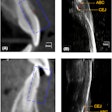
While magnetic resonance imaging (MRI) is often used for injuries of the knee and other body parts, researchers wanted to see if recent improvements in the modality meant it could be used to identify root cracks and fractures in teeth.
They compared the results from two imaging modalities: MRI and cone-beam CT (CBCT). The group stopped short of endorsing the modality for this task (Journal of Endodontics, May 2, 2019).
"Despite advantages of increased contrast and the absence of artifacts from radiodense materials in MRI, comparable measures of sensitivity and specificity (to limited field-of-view CBCT imaging) suggest MRI quality improvements are needed, specifically in image acquisition and postprocessing parameters," wrote the authors led by Tyler Schuurmans, DDS, from the division of endodontics at the University of Minnesota School of Dentistry in Minneapolis.
“Comparable measures of sensitivity and specificity (to limited field-of-view CBCT imaging) suggest MRI quality improvements are needed.”
Newer modalities
Newer 3D imaging modalities, such as limited field-of-view CBCT imaging, have been proposed as an aid to detect root cracks and fractures. However, the results from previous studies are mixed, and the modalities often require increased scan time and radiation dose to obtain an image, according to the authors.
MRI may offer an alternative modality to detect these cracks and fractures in teeth without ionizing radiation. The researchers wanted to see if they could determine the sensitivity and specificity of MRI and the current gold standard of CBCT imaging for detecting root cracks and fractures, compared with known crack and fracture status.
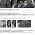
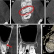
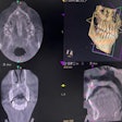
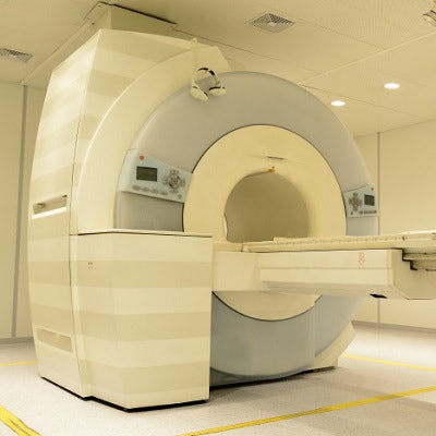
Dr. Schuurmans and colleagues matched 29 human adult teeth previously extracted after a clinical diagnosis of a root crack or fracture with the same number of control teeth. They used a 4-tesla, 90-cm bore, whole-body system (Magnex Scientific, Varian) and an ex vivo limited field-of-view CBCT device (CS 9000, Carestream Dental). A blinded, four-member panel rated the images.
The researchers reported that both modalities had comparably high specificity and poor sensitivity (see table below). They also noted that nearly half of the teeth had root canal filling materials identified on imaging.
| CBCT vs. MRI for identifying root cracks and fractures | ||
| CBCT | MRI | |
| Sensitivity | 0.59 | 0.59 |
| Specificity | 0.90 | 0.83 |
"MRI showed comparable measures of sensitivity and specificity for the detection of cracks/fractures in teeth in relation to limited field-of-view CBCT," the researchers wrote.
Future role?
While the researchers did not list any study limitations, they noted that the study was small. Future research should include a greater number of teeth or an artificially induced crack/fracture model to explore other possible measurements, such as crack/fracture width or extent, they wrote.
They concluded that they could not currently endorse the use of MRI, even though it provides "increased contrast and the absence of artifacts from radiodense materials."
However, "given the early stage of technology development, there may be a use for MRI in detecting cracks/fractures in teeth," the researchers added.
