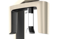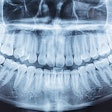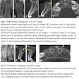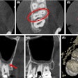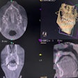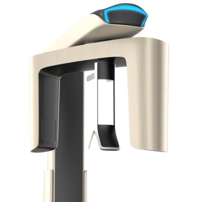
Accurately assessing furcation involvement in first molars can be difficult because of limited physical access, technology limitations, or measurement errors. Researchers of a new study wanted to see if cone-beam CT (CBCT) could help dentists overcome some of these limitations.
The researchers retrospectively compared the findings of CBCT images, periodontal probing, and radiographs in more than 80 patients with chronic periodontitis. They found that even an older version of a CBCT scanner provided more detailed clinical information on furcation involvement than the other techniques.
"This study validates that CBCT is a valuable tool in molar furcation assessment," wrote the study authors, led by Wenjian Zhang, DDS, PhD, of the department of diagnostic and biomedical sciences at the University of Texas School of Dentistry at Houston (BMC Oral Health, May 3, 2018).
First molar involvement
Furcation involvement refers to the condition when periodontal disease has caused bone resorption into the bifurcation or trifurcation of a multirooted tooth. But accurate determination of this bone loss is challenging. As CBCT may offer new clinical insight, the researchers wanted to compare the accuracy of clinical detection, intraoral radiography, and CBCT in assessing molar furcation.
The study included 83 patients (42 females) with chronic periodontitis. The researchers assessed furcation on maxillary and mandibular first molars. They used three methods to judge furcation involvement:
“This study validates that CBCT is a valuable tool in molar furcation assessment.”
- Periodontal examination by supervised predoctoral dental students and assessment of molar furcation involvement using the modified Glickman's classification
- Intraoral radiographs using a Focus wall-mounted unit (Instrumentarium Dental)
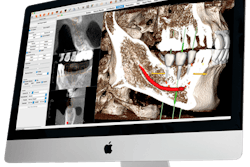
- CBCT images with a Kodak 9500 unit (Carestream Dental), and axial CBCT images reconstructed with Anatomage Invivo 5 software (Anatomage)
The modified Glickman's classification includes three classes:
- Class I, incipient or early stage of furcation involvement, means bone destruction is less than 2 mm into the furca.
- Class II is defined as horizontal bone destruction extending deeper than 2 mm but less than 6 mm into the furca.
- Class III is defined as horizontal bone destructions that result in a through-and-through tunnel.
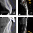
CBCT showed a higher correlation with clinical detection compared with intraoral radiography, especially at the distal palatal side of the maxillary first molar (p < 0.05), the researchers reported.
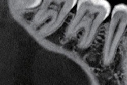
CBCT also provided the most accurate assessment of the three techniques, with bone loss measurement up to two decimal places in millimeters. The radiographs usually only detected the presence of furcation involvement in those patients falling into the Glickman class II and II categories.
The table below compares the clinical results from probing, radiographs, and CBCT:
| Average percentage of first molar furcation involvement found by modality | |||
| Modality | Radiographs | Periodontal probing | CBCT |
| Maxillary first molar | 28.2% | 29.6% | 32.6% |
| Mandibular first molar | 34% | 42.6% | 50.0% |
CBCT limitations
The authors listed several study limitations:
- The study was a retrospective investigation.
- There are interoperator variations in data collection, as the clinical detection was performed by different dental students under the supervision of a board-certified periodontist, and the results were later confirmed by supervising faculty.
- A relatively old model of CBCT unit was used.
In addition, CBCT itself has several limitations, including scatter, partial volume averaging, and beam-hardening artifacts, the authors noted. However, they recommended using CBCT for complicated cases when routine exams fail to provide adequate information for diagnosis or treatment planning.
"CBCT may be attempted with the smallest field-of-view possible and optimal exposure settings," they wrote.
