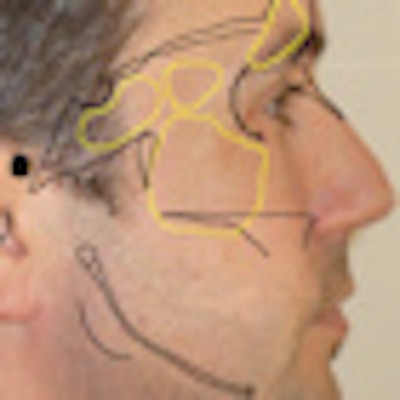
An interactive learning tool developed by researchers at the University of Queensland to enhance the learning of oral radiographic anatomy and interpretation received a warm welcome from dental students at the school who got to test-drive it (European Journal of Dental Education, February 17, 2011).
Studies reporting a high number of diagnostic errors made from radiographs suggest the need to improve the learning of radiographic interpretation in the dental curriculum, according to Camile Farah, BDSc, MDSc, PhD, an associate professor in oral medicine and oral pathology at the university, and colleagues noted.
Given that previous research has demonstrated a student preference for computer-assisted or digital technologies, "the purpose of this study was to develop an interactive digital tool and to determine whether it was more successful than a conventional radiology textbook in assisting dental students with the learning of radiographic anatomy," they wrote.
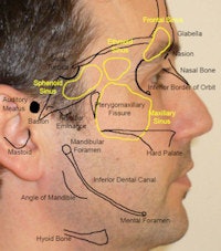
Click here to view PDF (3.4 MB)
Note: Due to the file size, the PDF make take extra time to download.
To create this tool, Dr. Farah and his colleagues made digital maps of anatomical hard- and soft-tissue structures using a digital photograph of a volunteer and the drawing functions of Adobe Photoshop. These maps were then superimposed on extraoral radiographs of the same volunteer. Ten transparency levels were then made of these superimposed images and exported into PowerPoint.
"Hence, the tool was essentially a PowerPoint presentation consisting of numerous slides of varying transparency of hard- and soft-tissue anatomy superimposed on the same radiographic image," they wrote.
The interactive function was the ability to scroll through these transparencies, allowing the addition or subtraction of soft-tissue structures from the hard-tissue structures displayed on the radiographs. Text and labels also were incorporated into the tool, indicating the various anatomical features general discernable in radiographs.
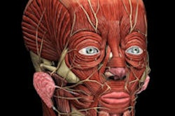
"The digital tools were developed as an easy resource for our students to be able to correlate soft- and hard-tissue anatomy in gross form and on radiographs," Dr. Farah told DrBicuspid.com. "We find commonly that students struggle to localize soft tissues on radiographs, particularly on panoramic radiographs."
3-pronged approach
The first phase of the study included 64 second-year undergraduate dental students attending the University of Queensland School of Dentistry in 2000. Second-year students were specifically invited because they had no prior learning in radiographic anatomy but did have some learning in head and neck anatomy as part of the dental curriculum.
The students were divided into two groups, and each student underwent a one-hour intervention phase involving the learning of radiographic anatomy using a conventional oral radiology textbook or the digital tool. Both resources were accessed online via the University of Queensland Online Learning Management System. All participants were then assessed on their understanding of radiography anatomy using 12 multiple-choice questions based on radiographic anatomy as seen on an orthopantomogram.
The participants then underwent a second one-hour intervention of learning, with each group using the other resource. They then answered another set of 12 multiple-choice questions to assess what they hard learned, and the results of both tests were compared. In addition, each student was asked to complete a qualitative questionnaire to assess their impressions regarding the effect of the digital tool on their learning of oral radiographic anatomy compared with the conventional oral radiology textbook.
In phase two, the study was repeated in exactly the same manner but with 24 fifth-year undergraduate dental students. Finally, in phase three, all 88 participants underwent assessment of their visual-spatial ability.
The results of the multiple-choice questions show that it did not matter whether the students used the textbook or digital tool in the first learning intervention phase (mean score of 51.3% for the textbook versus 50% for the digital tool), the researchers noted. In fact, the scores did not change significantly after the two groups repeated the learning intervention phase with the alternative resource.
However, the questionnaire results clearly indicated that the majority of students (94%) preferred using the digital tool rather than the textbook. In addition, all the participants (100%) found the digital tool easy to use, and the majority (85%) felt more confident learning with the digital tool than with the textbook.
Most notably, the researchers wrote, "the results show that a remarkable 95% of students felt the digital tool positively enhanced their learning of radiographic interpretation, with 97% agreement that it positively affected their performance."
Digital preference 'not surprising'
Only 21% of the students thought the digital tool alone was sufficient to learn radiographic anatomy, with the majority (88%) indicating the need for both resources. In addition, only 15% of the students felt that the textbook alone was sufficient to learn radiographic interpretation.
"With ongoing implementation of digital technologies across a variety of curricula, it is not surprising that the current generation of students shows immediate preference for computer-assisted learning and reference tools," Dr. Farah and his co-authors wrote.
However, they noted, the data indicate that while the digital tool positively affected how the students learned, it did not significantly affect what they learned.
“We would be happy to share the tool with other schools.”
— Camile Farah, BDSc, MDSc, PhD,
University of Queensland
"This suggests that the content of material learnt was consistent for all students; however, there was an obviously different impact of the methods used to deliver that content," they wrote. "Using the digital tool, when compared to the textbook, positively enhanced the learning process by enabling students to interact and better engage with the course material."
While previous studies comparing the learning outcome of conventional and digital resources have yielded similar results -- with a preference for digital -- Wisam Al-Rawi, BDS, MSc, an assistant professor in the department or oral diagnosis and radiology at Case Western Reserve University School of Dental Medicine, sees limitations to the approach used in the University of Queensland study.
Dr. Al-Rawi has developed two iPhone apps to help teach his students craniofacial anatomy that he says have been quite well-received.
"I would definitely say I got a very positive response from the students regarding using their iPhones to learn through an app versus a textbook," he told DrBicuspid.com. "They see textbooks as 'boring' instructional material. But the digital tool this study is talking about is merely different PowerPoint slides, and I honestly don't see any interactivity. It also still requires the student to sit in front of a computer to use it."
An iPhone app such as his iPanoramic, for example, allows zooming in and out, interactive hot spots, automatic multilingual support, and a quizzing module to enable students to periodically assess their progress. Plus it does not require a computer and is accessible through the iTunes store to millions of people all over the world, Dr. Al-Rawi noted.
"Basically you have your panoramic radiograph atlas in your pocket," he said. "That is very powerful!"
But Dr. Farah said he believes the university's digital learning tool is a good option in many situations.
"I do understand that other digital and more advanced software applications are available on the market, but the costs can sometimes be prohibitive," he said. "Our tools are made in PowerPoint and then converted to a PDF. This allows a small file size, and allows us to place the tool on learning management sites we employ in the school such as Blackboard. The tools then are available to all students as a learning resource. It makes access easy and reliable. We would be happy to share the tool (with some modifications to acknowledge the work that has gone into them) with other schools. We believe that other dental students can benefit from our simple, yet effective approach."
