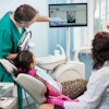
Interproximal decay, 25 years ago, when I was a dental student, was pretty cut and dry. The lesion, noted in a bitewing radiograph, was an indication of a cavity, and it needed to be drilled out and filled with a class II restoration. This meant that, if I showed the radiograph below to my clinical preceptor and suggested that we would have to do three fillings, my instructor would nod, give me the green light, and I would then commence the following:
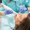
First, numb the patient with a needle. Drill a class II prep, removing decay, opening the contacts, and being careful not to nick the tissue or the adjacent teeth. (Something that, 25 years later, I’m still not perfect at.)
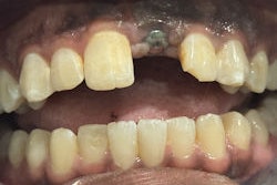
Place a matrix of some kind. Place a wedge, expecting to seal the gingival portion of the proximal box (which doesn’t always seal perfectly.)Figure 1: I don’t think I sealed that gingival margin.Images and captions courtesy of Dr. Ankur Gupta.
Place a sectional matrix ring, expecting to seal the buccal and lingual portion of the proximal box. (Something that, too, doesn’t always work out as planned.)
Selectively etch the beveled enamel (hoping that my etch doesn’t “splush” out the etch syringe, getting all over the dentin).
Figure 2: The etch may have “splushed” a bit here.
Scrub in the bonding agent for the manufacturer’s suggested amount of time. Fill the prep without voids, bubbles, overhangs, or flash, with some occlusal anatomy and recreation of the marginal ridge.
Sound easy?
It is for this reason that when I lecture to various groups throughout the country, I ask, “Do class IIs suck?” Most of the members of my audience happily raise their hands in agreement.
While we have all learned this particular series of steps as the gold standard in treating an interproximal lesion, is it possible that we have it wrong?
To start with, let’s discuss the nature of decay. Our enamel rods are held together with superglue. When we have sugar, the cariogenic bacteria that live in our mouths consume the sugar, release acid as a part of their metabolism, and that acid dissolves the superglue. This is called demineralization.
The reason that we don’t all need fillings even though most of us consume sugar is because a few hours later, the minerals in our saliva come along and “reglue” the enamel rods together, a process called remineralization.
If a person has an imbalance between remineralization and demineralization -- which could be due to a variety of factors -- they may start to lose some of those enamel rods and develop a cavity. What are those factors?
Not enough saliva
Saliva with inadequate mineralization potential
A high-sugar diet
Consumption of poorly timed sugar
An oral microbiome with excess cariogenic bacteria
Figure 3: Feel free to copy and modify this infographic for your own office. You can download the Canva image on my website bebetterseminars.com.
There are probably others, but let’s focus on the ones mentioned above. When I look at that list, I think about several potential solutions that don’t involve a drill, such as:
Diet counseling helps patients realize the effects of sugar and helps them to modify their consumption and modulate the timing of consumption.
Frequent application of fluoride, via chairside topical fluoride, or prescription-strength high-fluoride toothpastes and gels could bridge the mineralization gap if the patient’s saliva alone is not enough.
Curodont application to incipient lesions in the enamel can help remineralize the enamel via proprietary peptide-sequestering minerals from the patient’s own saliva, reversing the lesion without turning the tooth black.
Over-the-counter or prescription-strength salivary substitutes can be recommended when dry mouth is suspected.
Silver diamine fluoride effectively arrests (and can occasionally reverse) the progression of decay, even if some of that decay extends into the dentin.
That’s a lot of options, and while some dentists choose to use only a few of those options or none at all, there is no doubt that options exist between drill and do nothing.
Diagnostic innovation really is necessary to determine whether or not a lesion exists, along with the depth of that lesion. How are these lesions diagnosed? For most dentists, the diagnostic process includes two things: a scaler and an x-ray.
This process, in my opinion, is antiquated. It is difficult to orient a scaler in between teeth, and x-rays in general can sometimes be a nebulous and misleading collection of various shades of gray, highly dependent on the level of thoroughness from the clinician.
I would like to introduce three diagnostic innovations that we probably should be employing, but we may not know about.
The first diagnostic innovation is AI-supported radiographic interpretation
Rather than the discernment of various shades of gray resting on the shoulders of the clinician, who may or may not have adequate time in every different clinical scenario, decay is highlighted by the computer AI software itself, making it a bit more difficult to miss a lesion and highly documenting the degree of enamel or dentin penetration of the lesion.
Figure 4: Traditional bitewing radiography indicates clear decay on tooth #13, with some evidence of incipient lesions on #s 12, 14, 18, 19, 20, and 21. It is quite possible for me to miss one or more of these lesions on the lower teeth during busy times at the office.
Figure 5: AI radiographic interpretation helps highlight lesions and provides insight as to the extent of the decay.
The second diagnostic innovation is the pH evaluation of cariogenic bacteria inside a lesion
Figure 6: CaviSense toothpick cavity detectors.
Active decay will have a cavity full of cariogenic bacteria. Consumption of sugar will turn on the metabolic acid production in these bacteria. So, if a person were to chew on a sugary substrate, like a piece of candy, there would be a decrease in pH wherever an active cariogenic lesion exists. More can be learned about this by visiting Cavisense.com.
The third diagnostic innovation is transillumination of the interproximal space
Enamel possesses a certain translucency, allowing light to pass through without interruption. A cavity, on the other hand, creates an opacity (a shadow). Therefore, when illumination is applied buccolingually, the cavity is easy to identify. This is valuable for both the identification of dental caries but also for determining the extent of the decay.
The Catapult Product Evaluation Team, a group of dentists who apply new dental products and innovations into their already active and busy practices, was recently introduced to the DIAGNOCam by KaVo. This is a full-vision high-definition camera with three modes. The intraoral mode is similar to most intraoral camera features, but this camera has HD clarity. The two other modes, fluorescence and near-infrared transillumination, are geared more toward diagnosis. The fluorescence mode allows the practitioner to see caries, plaque, and occlusal attrition with greater clarity than with a traditional intraoral photo. Images in all three modalities can be captured simultaneously and displayed side by side.
Figure 7: Evaluating the marginal seal of an old resin filling. Transillumination shows some leakage, and the red fluorescence signal shows bacterial activity.
The transillumination mode, however, for me, was the game changer. The DiagnoCam’s special tip emits near-infrared light at the gingival level, from both the buccal and lingual aspects. The near-infrared light transmits through healthy enamel toward the camera, which has a corresponding bright, white appearance on the resulting image. Areas of carious or demineralized enamel scatter the light, resulting in a corresponding darker region.
Figure 8.
Using the same radiograph from above (Figure 8), I transilluminated the questionable areas, yielding highly accurate discernment of the extent and location of various cavities.
Figure 9: This is the interproximal space between teeth #s12 and 13. Notice that, upon learning the location of these interproximal lesions, the practitioner doesn’t have to prepare the lingual extent of the interproximal box.
Figure 10: This is the interproximal space between teeth #20 and #21. Notice that both lesions are incipient and do not extend into the dentin. This provided further confirmation that a traditional class II prep would not be appropriate.
The participating Catapult Evaluators were asked the following questions:
Was the DIAGNOcam helpful in providing early detection?
Did the DIAGNOcam provide a positive impact on identifying treatment opportunities?
Did it show enamel change that wasn’t visiblein the radiographs?
Did the DIAGNOcam highlight fractures?
Did the camera highlight secondary decay?
Did it make a strong positive impact on how patients understood their condition?
The responses to this product review were incredible. Over 85% of survey respondents remarked positively on the questions above. Evaluators were most impressed with the ability of the DIAGNOcam to provide visual confirmation of pathology that simply was not present in the radiographs.
One evaluator remarked, “There was a lingual crack on #3 that was completely missed on the radiograph, and was very difficult to see visually with an intraoral exam. Fluorescence and transillumination made the fracture so obvious. This was helpful both for me and for the patient.”
One evaluator remarked, “I could finally see recurrent decay in three dimensions instead of guessing from the films.” While another evaluator said, “Radiographs pick up the lesion about half the time, and DIAGNOcam showed me enamel changes that I could not see any other way.”
I personally found the DIAGNOcam to be incredibly helpful, especially when trying to determine the level of invasiveness in a traditional cavity prep. Because I was able to discern the buccolingual location of the lesion, it allowed my prep to be the least invasive, which I appreciated.
Patient communication and education were similarly outstanding. For some reason, while patients don’t quite see what you are talking about when you share radiographs, they seem to have a greater level of understanding while looking at transillumination between the teeth.
Lastly, there is a growing number of patients and parents who are uncomfortable with traditional healthcare interventions, including fluoride and radiographs. While I do not particularly agree with their hesitation toward those features of our dental repertoire, it is important for me to meet the patients where they are and try to accommodate their wishes. Providing an innovative and accurate means of determining interproximal decay, without the use of any radiation, is very helpful in these situations.
What ultimately set DIAGNOcam apart was how its technology fit into a modern, prevention-focused practice. Its innovative imaging approach complements radiographs and AI rather than competing with them, giving us another layer of diagnostic confidence. Taken together, these factors support DIAGNOcam earning the Catapult Vote of Confidence™.
Editor's note: Learn more about DIAGNOcam from Dr. Gupta below.
The comments and observations expressed herein do not necessarily reflect the opinions of DrBicuspid.com, nor should they be construed as an endorsement or admonishment of any particular idea, vendor, or organization.
Dr. Ankur A. Gupta graduated from the University of Michigan School of Dentistry. After completing a one-year general practice residency in Cleveland, he and his partner, Dr. Nisha Gupta, started North Ridgeville Family Dentistry in Ohio. Gupta is a member of the ADA, the Greater Cleveland Dental Society, the Ohio Dental Association, the ADA Success Speaker Corp, and he is a board member and speaker for Catapult Education’s Speaker Bureau. Gupta is an Academy of General Dentistry Program Approval for Continuing Education-certified provider.
 Dr. Ankur Gupta.
 Figure 1: I don’t think I sealed that gingival margin.Images and captions courtesy of Dr. Ankur Gupta.
 Figure 2: The etch may have “splushed” a bit here.
 Figure 3: Feel free to copy and modify this infographic for your own office. You can download the Canva image on my website bebetterseminars.com.
 Figure 4: Traditional bitewing radiography indicates clear decay on tooth #13, with some evidence of incipient lesions on #s 12, 14, 18, 19, 20, and 21. It is quite possible for me to miss one or more of these lesions on the lower teeth during busy times at the office.
 Figure 5: AI radiographic interpretation helps highlight lesions and provides insight as to the extent of the decay.
 Figure 5: AI radiographic interpretation helps highlight lesions and provides insight as to the extent of the decay.
 Figure 6: CaviSense toothpick cavity detectors.
 Figure 7: Evaluating the marginal seal of an old resin filling. Transillumination shows some leakage, and the red fluorescence signal shows bacterial activity.
 Figure 8.
 Figure 9: This is the interproximal space between teeth #s12 and 13. Notice that, upon learning the location of these interproximal lesions, the practitioner doesn’t have to prepare the lingual extent of the interproximal box.
 Figure 10: This is the interproximal space between teeth #20 and #21. Notice that both lesions are incipient and do not extend into the dentin. This provided further confirmation that a traditional class II prep would not be appropriate.