Swept-source optical coherence tomography (SS-OCT) offers a cross-sectional, high-resolution view of teeth, allowing dentists to diagnose caries, cracks, and more without exposing patients to radiation, according to an article published in the November issue of Japanese Dental Science Review.
The imaging technology can be used on infants, young children, and pregnant women without exposing them to x-ray radiation, and, as the technology evolves, it may prove useful in the diagnosis of other conditions, such as oral soft-tissue diseases and periodontal disease, the authors wrote.
"In dentistry, OCT imaging is effective for the diagnosis of dental caries, tooth cracks, and aging changes of tooth structures such as [noncarious cervical lesions (NCCL)] and occlusal tooth wear," wrote the group, led by Dr. Yasushi Shimada from the Graduate School of Medicine, Dentistry, and Pharmaceutical Sciences at Okayama University in Japan (Jpn Dent Sci Rev, November 2020, Vol. 56:1, pp. 109-118).
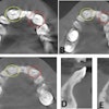
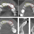
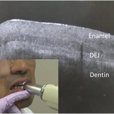
 SS-OCT was used on the anterior tooth to provide real-time cross-sectional imaging. The right side of the image shows enamel and dentin, as well as the dentin-enamel junction appearing as a border between the two structures. All images courtesy of Shimada et al. Licensed under CC BY-NC 4.0.
More precision
Optical coherence tomography is an imaging technique that creates cross-sectional images of translucent or semitranslucent biological structures, like teeth, with a microscopic level of resolution. Unlike with x-rays, OCT uses light to create images, so there is no risk of radiation exposure.
Early OCT systems used time-domain detection, which is when echo time delays of light are determined by measuring the interference signal as a function of time, while scanning the optical path length of the reference arm. The technology is ever-changing. The latest version is SS-OCT, and, with it, image resolution and speed have improved significantly. With SS-OCT, a tunable laser sweeps near-infrared wavelength light, providing real-time imaging, the authors wrote.
A clear, internal view
When this approach is used, sound enamel becomes nearly clear at the OCT wavelength range. It is easy to differentiate between enamel and dentin at the dentin-enamel junction because it looks like a dark border. Demineralized enamel and dentin show up as bright zones. This is due to the formation of many microporosities where the backscatter of imaging signals is increased, according to the authors.
Using SS-OCT, the upper margin of a cavity reflects the signal, appearing as a distinct bright border in cases of cavitated caries at the interproximal or occlusal hidden zone. This imaging approach can also determine the crack penetration depth of a tooth even when the fracture extends beyond the dentin-enamel junction, they wrote.
 SS-OCT images of occlusal caries (A1, B1) and corresponding histological view after cross-sectioning (A2, B2). Enamel demineralization at the occlusal fissure is imaged as increased brightness within the depth of the dentin-enamel junction (arrow 1). The presence of hidden dentin caries is depicted in SS-OCT as backscattering signal from the lesion boundary (B1). Lesion expands along the dentin-enamel junction, appearing as a bright line (arrow 2).
Future uses
Due to its high degree of sensitivity and specificity, SS-OCT is effective for detecting caries, cracks, noncarious cervical lesions, and occlusal tooth wear. As the technology continues to advance, it likely could be used in other diagnoses, according to the authors.
"Further technological evolution will allow [one] to utilize the OCT imaging for diagnosing periodontal disease and oral soft-tissue diseases," they wrote.

