SAN FRANCISCO - To detect cracks in ceramic restorations, swept-source optical coherence tomography (SS OCT) may be a better option than 2D microscopy, according to new research presented Wednesday at the International Association for Dental Research (IADR) annual meeting.
 John Sorensen, DMD, PhD, presented his OCT research at IADR 2017.
Researchers from the University of Washington and Tokyo Medical and Dental University compared current methods for evaluating cracks in ceramics against newer OCT techniques. They found that, at least for lithium disilicate ceramic, OCT outperformed optical microscopy at detecting and evaluating cracks.
"It allows us to really evaluate in three dimensions -- track those cracks down and observe them in a way we've never been able to do before," said presenter John Sorensen, DMD, PhD, a professor at the University of Washington School of Dentistry and the associate dean for clinics and director of research for the graduate prosthodontics program.
New uses for OCT
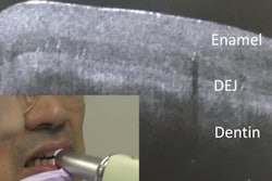
Although OCT has been used in ophthalmology for more than a decade, it is a relatively new technology for dental and other medical research. Dr. Sorensen and colleagues wondered if swept-source optical coherence tomography might be a reliable 3D imaging technique for detecting cracks in ceramic onlays, since traditional imaging methods, including optical microscopy, lack sensitivity.
“This is a very powerful tool for 3D detection”
The researchers began by cementing 20 lithium disilicate ceramic onlays to molars with no occlusal enamel. They broke the onlays into four groups of five onlays with varying thickness levels: 0.3 mm, 0.5 mm, 0.8 mm, and 1.5 mm. After subjecting the onlays to simulated chewing forces, they then imaged the onlays using both optical microscopy and OCT.
The OCT technique detected more in-depth cracks and interfacial debonding in the ceramic onlays than optical microscopy. As a result, the researchers are optimistic about the future of OCT in dentistry.
"SS OCT was remarkably more sensitive for detection of cracks than optical microscopy," Dr. Sorensen said. "This is a very powerful tool for 3D detection, dynamic visualization, and actually quantification of cracks and interfacial gaps in cemented ceramic restorations."
OCT limitations
While OCT does seem promising for dentistry, the technology still has a number of shortcomings. For instance, the technique doesn't work well with more acidic ceramics, which is why the researchers used lithium disilicate ceramic. Additionally, the researchers had to exclude the outer 20 microns of the OCT image because of scatter.
Nevertheless, Dr. Sorensen is optimistic that OCT may be a valuable tool for helping detect cracks in some ceramic restorations.
"This is a very exciting area," he told DrBicuspid.com. "It works very well in dental hard tissues. ... It lends itself very well to some of the ceramics we have available today."
