What role can imaging play in determining the diagnosis of temporomandibular disorders (TMDs) in patients with suspected cases?
Researchers from Switzerland sought to build on previous studies to determine if imaging findings lead practitioners to believe patients have temporomandibular pain when actually there is none.
"In individuals with manifest[ed] or suspected TMDs, clinical decision-making as well as TMD-related prognostic statements of dentists are primarily based on somatic factors, such as the presence of temporomandibular pain, while psychosocial variables do not appear to play an important role, if any," lead study author Jens Türp, DDS, and colleagues wrote (BMC Oral Health, November 17, 2016).
Dr. Türp is a professor in the department of reconstructive dentistry and temporomandibular disorders at the University Center for Dental Medicine Basel at the University of Basel.
Gold standard
When determining if a patient has a TMD, practitioners often look at three key factors:
- The position of articular disk relative to the mandibular condyle
- The location of the condyle relative to the temporal joint surfaces
- The depth of the glenoid fossa of the temporomandibular joints (TMJs)
The researchers wanted to determine if these variables were actually prevalent in patients with TMDs based on two study cohorts (patients born in 1930-1932 and those born in 1950-1952). They used imaging studies to reinterpret the clinical significance of the findings.
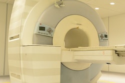
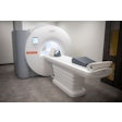
The researchers used magnetic resonance imaging (MRI) in their study, which they called the gold standard for imaging soft tissues. They also noted that in the TMJ area, MRI is the imaging modality of choice to investigate disk position and optimize depiction of the condylar cartilage.
The researchers examined existing MRI scans taken in 2005 and 2006 of the TMJs of 72 patients. The condylar position at closed jaw was calculated and a "textbook-like" disk position at closed jaw was distinguished from an anterior location. The researchers assessed the TMJ morphology of the temporal joint surfaces at open jaw by measuring the depth of the glenoid fossa.
There were 139 MRIs of closed and 130 MRIs of open jaw positions available for evaluation. For each patient, one slice of the sagittal views -- the slice located in the middle of the long axis of the condyle -- was selected from the open and closed positions for assessing both the mandibular condyle and the articular disk.
The researchers found that only 68 of 139 (48.9%) condyles were located centrically, whereas 71 (51.1%) were in an eccentric position. They also found that in 75% of the TMJs, the most frequent position of the articular disk corresponded to the one described in textbooks, which means that about 25% of these disks were anteriorly displaced. The frequency of this anterior displacement was twice as high in women as in men (34.5% for women and 17.1% for men).
They also found a significant difference in glenoid fossa depth in men and women and in younger and older patients (see table below).
| Glenoid fossa depth in younger and older patients | |
| Year born | Depth (mm) |
| 1930 (women) | 6.1 |
| 1950 (women) | 6.9 |
| 1930 (men) | 6.3 |
| 1950 (men) | 7.8 |
Reinterpretation needed
“We intend to protect nonpatients against medicalization, overdiagnosis, and unnecessary therapy.”
The researchers did not consider coronal views in this study, which might be considered a limitation, according to the authors. They also noted that while cone-beam CT might be a better modality for investigating hard tissues, such as condylar position and osseous changes, these scans "would not have been allowed by the ethical committee to be used," they wrote.
The authors concluded that reinterpretation of the significance and labeling of imaging findings and derived diagnoses is called for "because [these diagnoses] may insinuate pathology which usually does not exist." They recommended that expressions such as "condylar displacement" or "eccentric position" for describing a noncentric condylar position should be avoided.
"By doing so, we intend to protect nonpatients against medicalization, overdiagnosis, and unnecessary therapy," the authors wrote.
