Radiographic film is superior to phosphor-plate digital panoramic images for imaging low-contrast structures, according to a new study in Dentomaxillofacial Radiology (October 2010, Vol. 39:7, pp. 424-430).
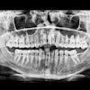
Researchers from Ege University in Turkey compared the quality of clinical images obtained with a storage phosphor plate (SPP)-based digital system versus a conventional film-based panoramic system in visualizing various anatomical structures. They also evaluated the effect of various processing algorithms on image interpretation.
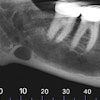
They took panoramic radiographs in 42 patients using both the film-based system and phosphor-plate system. The phosphor-plate images were treated with shadow, sharpen, negative, and exponential filters. Four observers evaluated all the radiographs and digital images to determine the visibility of anatomical structures with various radiodensities and overall image quality.
The observers found no statistically significant difference between film and unfiltered digital images except for low-contrast structures (p > 0.05), the researchers reported. Film images were preferred for the visibility of low-contrast structures (p < 0.05), and best overall image quality was obtained with sharpened images (p < 0.05) followed by films and unfiltered digital images. Among all filtered images, sharpened ones received the highest ratings for the visibility of all anatomical structures (p < 0.05).
"Film and unfiltered SPP-based panoramic images performed equally well in terms of overall quality," the researchers concluded. "However, films were best for the perception of low-contrast structures."
The sharpening filter may be recommended for enhancing SPP panoramic images to improve the visual perception of most of the anatomical structures and also overall quality, they added.
Copyright © 2010 DrBicuspid.com