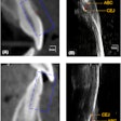
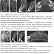
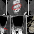
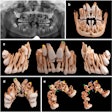
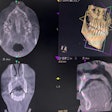
Combining the 3D imaging capabilities of cone-beam CT (CBCT) with a cephalometric simplified protocol can overcome the limitations of conventional 2D cephalograms and improve diagnoses, according to researchers from the University of Milan (Progress in Orthodontics, May 11, 2010).
Cephalograms have inherent limitations as a result of distortion, super imposition, and differential magnification of the craniofacial complex, which can lead to errors of identification and reduced measurement accuracy, the researchers noted.
To determine whether standard cephalograms could be improved by combining this 2D imaging technique with CBCT, they assessed cephalometric 2D and 3D measurements of the volume and centroid of the maxilla and mandible in 10 clinical cases.
With a few exceptions, they found that the linear and angular cephalometric measurements obtained from CBCT and conventional cephalograms did not differ statistically (p > 0.01). There was a correlation between the variation in the skeletal malocclusion and growth direction of the jaws, and the variation in the spatial position (x, y, z) of the centroids and their volumes (p < 0.01).
However, the 3D cephalometric analysis was easier to interpret than 2D cephalometric analysis, the researchers noted. In addition, the angular and linear measurements detected on 3D become "real," and the automatic measurements made by the computer drastically reduced human error, yielding a more reliable, reproducible, and repeatable diagnosis, they concluded.
Copyright © 2010 DrBicuspid.com
