While lateral radiographs provide "reliable evaluation" of the quantity of bone in preoperative diagnosis of palatal implants, additional 3D imaging is only required in rare cases of borderline dimensions, according to a new study in Clinical Implant Dentistry and Related Research (February 3, 2010).
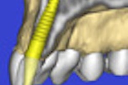
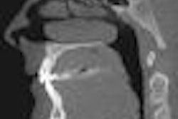
Researchers from the department of orthodontics at the University Medical Center Mainz set out to evaluate the diagnostic value of lateral radiographs in preoperative diagnostics for orthodontic anchorage implants, and whether CT or cone-beam CT (CBCT) are necessary as additional preoperative diagnostic tools in such cases.
They reviewed all patients who presented for insertion of a palatal implant between January 2003 and December 2007 at University Hospital Mainz. On the basis of lateral radiographs, the palatal bone was assessed as:
- Group A: sufficient (bone height > 4 mm in the implant axis)
- Group B: ambiguous
- Group C: insufficient (bone height < 4 mm in the implant axis)
In Group A the surgical insertion procedure was performed without further radiological investigation. Group C required other types of anchorage. In cases of an ambiguous bone situation (Group B, two patients), CT and CBCT were also performed.
A total of 105 patients were screened during the observation period. Fourteen patients opted for alternative treatment, leaving 91 patients for final evaluation. In 89 patients (98%), the lateral radiographs showed sufficient bone in the vertical dimension. In all of these cases, the availability of sufficient bone was confirmed intraoperatively.
"Lateral radiographs permit correct and reliable evaluation of the quantity of bone in preoperative diagnosis of palatal implants," the researchers concluded. "Additional imaging (CT or CBCT) is only required in rare cases of borderline dimensions."
Copyright © 2010 DrBicuspid.com