Editor's note: Allan Farman's column, Talking Pictures, appears regularly on the DrBicuspid.com advice and opinion page, Second Opinion.
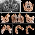
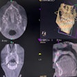
Without a doubt, 3D images from dedicated maxillofacial cone-beam computed tomographic systems will revolutionize the way advanced dental procedures are conducted. This is why sales of the cone-beam computed tomography (CBCT) systems continue to be strong world wide and why new competitors are continually entering the marketplace.
In a statement issued by the American Academy of Oral and Maxillofacial Radiology (AAOMR) this month, the AAOMR embraced the introduction of CBCT as a major advancement in the dental imaging armamentarium. However, the academy also voiced several cautions; namely, that the practitioner should apply imaging procedures based on considerations of patient radiograph selection criteria, dose optimization, technical proficiency, and assessed diagnostic or treatment needs.
The AAOMR believes that CBCT examinations should be performed only for valid diagnostic or treatment reasons and with the minimum exposure necessary for adequate image quality. In addition, it should be performed only by an appropriately licensed practitioner (or certified radiologic operator under supervision of a licensed practitioner) with the necessary training. In states where CBCT is considered to be a medical device, a dental assistant could conceivably not be credentialed to make a CBCT exposure. Further, documentary evidence should be provided to demonstrate the diagnostic or treatment guidance need of the CBCT examination.
There may be a misconception among some practitioners that the user has no responsibility for radiologic findings beyond those needed for a specific task (e.g., implant treatment planning). According to the AAOMR, this assumption is erroneous. To support the diagnostic necessity of the procedure and facilitate patient understanding, they believe it is desirable that a separate patient consent be obtained for the CBCT procedure prior to imaging.
The AAOMR also believes that practitioners should undergo specific training to perform CBCT examinations successfully. The practitioner who operates a CBCT unit or requests a CBCT study needs to examine the entire image dataset and have a thorough knowledge of CT anatomy of the entire acquired volume, anatomic variations, and observation of abnormalities and disease processes. However, qualified oral and maxillofacial radiologists may be able to assist diagnostically when practitioners are unwilling to accept the responsibility to review the whole exposed tissue volume.
Without a doubt, 3D imaging, including CBCT, will become the standard of care for many procedures in dental and maxillofacial practice. This represents a paradigm shift in dental diagnostic imaging, and it will take time for dental education to catch up. It also means that practitioners who graduated from dental school in pre-CBCT days will need to have their knowledge "retread" through continuing education and training programs.
The comments and observations expressed herein do not necessarily reflect the opinions of DrBicuspid.com, nor should they be construed as an endorsement or admonishment of any particular idea, vendor, or organization.
Copyright © 2008 DrBicuspid.com
