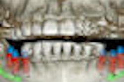
The growing use of cone-beam CT (CBCT) for diagnostic and treatment planning applications in dentistry has prompted a parallel body of discourse on the legal liabilities associated with 3D image interpretation.
These discussions have, in turn, prompted a number of reports of incidental findings in dental cone-beam CT images. In a study in the Journal of Implant & Advanced Clinical Dentistry, for example, a team of researchers from New York sent cone-beam CT data from 500 consecutive patients to nine dental imaging centers for review (April 2010, Vol. 2:3, pp. 87-91).
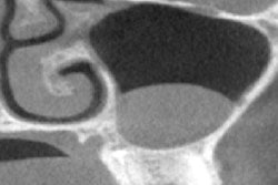
Of the 500 patients, 204 were referred exclusively for maxillary studies, and 79 were referred for both maxillary and mandibular studies. Of the 283 evaluated maxillary sinuses, incidental findings were found 76.3% of the time, and other incidental findings -- including impacted teeth, retained root tips, and bone grafts -- were found in 69.3% of the scans. The most frequent incidental finding was periapical radiolucencies (229/500).
"While dentists embrace 3D technology, they need to become familiar with normal and abnormal landmarks," the authors wrote. "In addition, they need to be prepared to identify incidental findings and inform their patients as to the existence of these findings and their clinical relevance."
AAOMR presentations
Two presentations at the recent American Academy of Oral and Maxillofacial Radiology (AAOMR) lend further support to these recommendations.
Most dentists who are not radiologists are not familiar with interpreting anatomical structures and/or pathosis outside their areas of primary interest, noted Veeratrishul Allareddy, BDS, an assistant clinical professor at the University of Iowa. As part of his 2009 master's thesis project, he had an oral and maxillofacial radiologist examine 1,000 cone-beam CT images to determine the number and nature of incidental findings, and bring this issue to the attention of general practitioners.
"Often they are not aware that if they order a radiograph, they are responsible for all findings on it," Dr. Allareddy said during his AAOMR presentation.
The images were acquired on an i-CAT cone-beam CT system (Imaging Sciences International) at a field-of-view of 13 cm and a 0.3-mm thickness in 8.5 seconds. The patients had been referred for the scans for various reasons, including bone evaluation for implants, impaction localization, orthodontic records, and supernumerary teeth localization.
Of the 1,000 scans reviewed, only 57 had no osseous pathosis or incidental findings, and 943 showed findings in the primary regions of interest and/or outside the regions of interest. More than 70 different conditions were visualized in the scans, both in and outside the areas of interest.
The most critical incidental findings, according to Dr. Allareddy, were three patients with malignant neoplasms that had previously not been detected.
"Once you order a cone-beam CT scan, make sure it is read by a radiologist," he concluded.
In a separate presentation, Nadia Yousefinejad of the University of Medicine and Dentistry of New Jersey shared findings of a cross-sectional study of reports from 427 cone-beam CT scans. The scans were submitted to the faculty practice of an oral radiologist certified by the American Board of Oral and Maxillofacial Radiology for consultation. Significant incidental findings were categorized based on the radiographic impressions that were noted in the reports. The initial reason for obtaining each scan was also recorded and analyzed.
The most frequent reasons for the scans were implant assessment and orthodontic diagnosis and treatment planning. Common incidental findings included impacted teeth, anatomic variations, inflammatory lesions, and sinus disease.
"Newer, small-volume cone-beam CT machines, application of selection criteria, and improved radiographic knowledge on the part of the practitioner may aid in appropriate future management of the data obtained from cone-beam CT scans," the researchers concluded.
Liability concerns
|
Sending images out for review Companies such as Orbit Imaging, Dimensions Imaging, 3-D Diagnostix, and 360imaging contract with oral and maxillofacial radiologists to read image volumes for unsuspected pathologies in cone-beam CT scans. The cost averages between $60 and $120 per scan. |
The AAOMR recommends that dentists should be competent to identify abnormalities and suspicious areas of pathosis in the entire cone-beam CT scan or refer the images to a specialist for final interpretation, according to Edwin Zinman, DDS, JDS, a San Francisco attorney who specializes in dental law (Journal of the California Dental Association, January 2010, Vol. 38:1, pp. 49-56).
"Don't be fooled or lulled into a false sense of security just because of the low statistical likelihood of overlooking uncommon or rare lesions such as neoplasms or other tumors on cone-beam CT images," Dr. Zinman stated in an e-mail to DrBicuspid.com. "There is no average patient. If a lesion is present on a cone-beam CT scan, then for that individual patient their statistic is 100%. Consequently, 100% of cone-beam CT scans must be diagnosed for pathosis 100% of the time by viewing 100% of cone-beam CT scanned images."
The standard of care requires reasonable care, which means care based upon reason, he added. "As dental scientists, this reason is scientific reasoning based upon peer-reviewed research studies," he said.
For all reasonable practitioners, cone-beam CT should become part of the armamentarium "for judicious selection," Dr. Zinman said.
"As with all paradigm shifts, it will be both used and abused," he concluded.
Copyright © 2010 DrBicuspid.com
