Cone-beam CT is better for preoperative radiographic evaluation of complicated impacted lower third molars than other imaging modalities, according to new research from the University of Helsinki (Oral Surgery, Oral Medicine, Oral Pathology, Oral Radiology, and Endodontology, February 2010, Vol. 109:2, pp. 276-284).
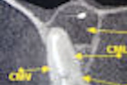
The researchers compared the reliability of cone-beam CT with that of panoramic radiography, multiprojection narrow-beam radiography (MNBR), and cross-sectional tomography in preoperative radiographic determination of the number of roots of lower third molars and their relationship to the inferior alveolar canal (IAC).
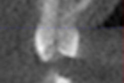
Forty-two teeth were clinically studied and imaged using all four modalities. Statistical analysis was used to compare the diagnoses of two trained oral radiologists and the radiologic diagnoses with the findings at operation.
Cone-beam CT revealed the number of roots of teeth more reliably than panoramic radiographs, the researchers noted.
"Cone-beam CT examination was highly reliable in locating the IAC, whereas MNBR was unreliable and cross-sectional tomography fell between the two," they wrote. In addition, with cross-sectional tomography, the IAC was noninterpretable in one-third of the cases.
Copyright © 2010 DrBicuspid.com