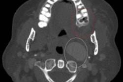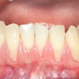
Lesions appearing on radiographs with gum swelling are often misdiagnosed as lateral periodontal cysts instead of odontogenic keratocysts, which are more aggressive. The study findings were published on December 16 in the Journal of Endodontics.
Both types of lesions can present with similar clinical and imaging appearances; however, the rare but benign keratocyst is a locally aggressive developmental cyst with a high rate of recurrence. Therefore, a diagnosis needs to be confirmed with histological findings, the authors wrote.
"A biopsy should be considered in all cases to establish the correct pathologic diagnosis and treatment course," wrote the group, led by Dr. Janina Golob Deeb, an associate professor in the periodontics department at the Virginia Commonwealth University School of Dentistry.
It can be challenging to provide an accurate diagnosis for lesions in areas of overlapping regional, clinical, and imaging characteristics, the authors noted. Careful radiological, clinical, and histological exams are needed to treat lesions appropriately. This is particularly important with odontogenic keratocysts: Due to the deeply seated daughter cells associated with odontogenic keratocysts, the lesion has a high chance of recurrence if not treated properly.
In the current study, the researchers retrospectively analyzed pathology reports that included 79,257 biopsies. Deeb and colleagues used the data to assess the number of lateral periodontal cyst and odontogenic keratocyst cases. They examined the alignment of clinical and radiographic diagnosis to histologic findings and anatomical location and identified the number of preoperatively misdiagnosed cases of odontogenic keratocyst.
Among the biopsies, 184 were diagnosed as lateral periodontal cysts and 742 as odontogenic keratocysts. For all preoperatively diagnosed lateral periodontal cysts, the clinical and histological diagnoses aligned. However, 182 (25%) of the 742 cases of odontogenic keratocyst were submitted with a clinical misdiagnosis of lateral periodontal cyst, the authors wrote.
Most likely, these cases were misdiagnosed based on their radiographic and clinical presentations, as well as the treating clinician's judgment. The misdiagnosed lesions were often located in the maxillary and mandibular anterior and premolar regions, they wrote.
Due to the significant number of potentially misdiagnosed lesions, clinicians should conduct a thorough review of differential diagnoses when a patient presents with a cystic lesion in the interbicuspid area, Deeb and colleagues wrote.
"This underscores the importance of histologic examination of the removed tissue to establish a proper diagnosis and treatment plan," they concluded.


