An oral surgeon diagnosed and treated a teen with a rare vascular tumor in her mouth after a dentist and periodontist improperly treated her for periodontitis for two years. Details of the case were published on November 6 in BMC Oral Health.
After tissue samples were collected and analyzed, the patient was diagnosed with the rare malignant vascular tumor epithelioid hemangioendothelioma (EHE), which can mimic periodontal disease. This case is one of about 40 reported cases of EHE occurring in the mouth, and it highlights the importance of being aware of this unusual tumor, according to the authors.
"This rare tumor must be included in differential diagnoses of periodontal pathologies to perform histomorphological examination in a timely manner, which could lead to correct diagnosis and adequate treatment," wrote the group, led by Dr. Gintaras Januzis from the department of maxillofacial surgery at the Lithuanian University of Health Sciences in Kaunas.
Epithelioid hemangioendotheliomas are rare tumors that account for up to 1% of all vascular tumors. They are most often found in the liver and lungs and are rarely found in the mouth. This type of tumor grows slowly, and its symptoms imitate those of chronic inflammation, such as gum disease, which makes it more difficult to diagnose.
An 18-year-old woman
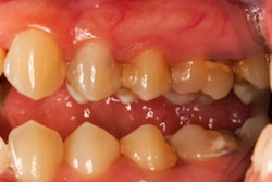
A healthy woman went to the dentist with gingival problems around her upper right premolar. She was diagnosed with marginal periodontitis and was referred to a periodontist for root scaling. For two years, the woman underwent periodontal treatments while taking anti-inflammatory medications. She also had a root canal on her second premolar. Eventually, she was referred to an oral surgeon because her condition was not resolving.
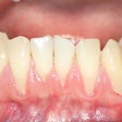
When she visited the surgeon, her only complaint was gingival recession in the palatal area of the upper right side of her teeth. An exam revealed that her canine and both premolars had second-grade mobility. The probing depth of teeth #13, #14, and #15 was less than 3 mm on the buccal side and 5 mm at the palatal side. Gum recession left the first premolar and canine exposed, and the exposed root surface of the second premolar was covered with plaque. A 3D computed tomography (CT) scan was taken of her jaw. It showed bone destruction in the defect area that reached the maxillary sinus, where mucosa was locally thickened, according to the authors.
Didn't add up
The oral surgeon was confused by the chronic lesion that had no known cause and an odd localization. The clinical findings were not characteristic of cancerous tumors. Therefore, the surgeon and the patient decided that her #13, #14, and #15 teeth would be removed along with altered soft tissue. The collected tissue was sent to the lab for evaluation, the authors noted.
The surgeon diagnosed her with epithelioid hemangioendothelioma based on the bony destruction seen on x-rays, as well as the accumulation of tumor cells assessed by immunohistochemical staining.
Reconstruction using an allogenic bone transplant began 12 months after the tumor was removed. After 31 months, the patient had shown no clinical or radiographic signs of relapse, the authors wrote.
In the report, the authors noted some limitations, including the fact that the clinical presentation did not raise immediate concerns of a neoplastic lesion. Therefore, an initial biopsy was not performed, they noted.
Avoiding misdiagnosis
The patient lost three teeth due to the lesion expanding before the correct diagnosis was made. This case illustrates the importance of dentists becoming familiar with the rare tumor to avoid misdiagnosis in the future.
"All of this could have been prevented by timely performed histomorphological examination, which could have led to correct diagnosis and adequate treatment," they wrote.


