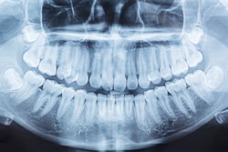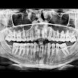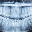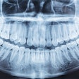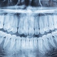
Since nearly 3 in 4 healthy kids may present with at least one dental abnormality or pathology, dentists should order panoramic x-rays at ages 9, 12, and 15 to detect them. The study was published on June 17 in the International Journal of Paediatric Dentistry.
The study validates the diagnostic efficacy of panoramic x-rays for diagnosing developmental dental anomalies and pathologies (DDAPs) in pediatric dental patients, the authors wrote.
“Age can be a predictor for defining frequency of PRs (panoramic radiographs) to detect DDAP in children,” wrote the authors, led by Dr. Gary Schulman of the University of Connecticut School of Dental Medicine in Farmington.
Since minimizing radiation exposure is vital in developing children, the ADA and the American Academy of Pediatric Dentistry recommend that clinicians conduct a thorough review of the patient's history, clinical examination, prior imaging, caries risk assessment, and medical and dental needs prior to prescribing x-rays.
Though dental x-rays are important for the diagnosis and treatment of pediatric patients, there are no objective, evidence-based guidelines regarding the frequency of panoramic radiographs in pediatric patients. The age-based evaluation of x-rays for dental pathologies and abnormalities will provide objective data for dentists to determine how often children should undergo imaging, according to the study.
To evaluate the age-based prevalence of DDAPs on panoramic x-rays and to determine when the appropriate age is to detect these conditions, the authors conducted an observational cohort study. X-rays from 581 patients between the ages of 6 and 19 were reviewed to identify anomalies of size, shape, position, structure, and other developmental anomalies and pathologies of the face and neck region, they wrote.
Of the children, 411 (74%) had at least one anomaly. Of the anomalies, 12% were shape-related; 17% were number-related; 28% were positional-related; and 63% were other dental anomalies and pathologies, including condylar erosion, vascular calcifications, and fibrous dysplasia, the authors wrote.
The optimal Youden index cutoff, which measures the effectiveness of a diagnostic marker and allows for the optimal cutoff point for the marker, for any anomaly was age 9, with an area under the curve (AUC) of 0.73 (95% confidence interval [CI], 0.68 to 0.77). After the first cutoff at age 9, the next was at age 12 (AUC: 0.65; 95% CI, 0.59 to 0.71), and age 15 was the optimal secondary cutoff for both number and positional anomalies (AUC: 0.57; 95% CI, 0.48 to 0.65; AUC: 0.64; 95%CI, 0.58 to 0.71, respectively), they wrote.
Nevertheless, the study had multiple limitations, including that the dental age may not have matched the chronological age for some of the patients, the authors wrote. In the future, studies should be conducted to document the incidence of dental anomalies and pathologies based on chronological and dental ages to investigate the correlation between the abnormalities and diseases and their linked findings in pediatric patients, they wrote.
“(For early and timely detection), PRs should be prescribed at ages 9, 12, and 15 years for the diagnosis of DDAP (developmental dental anomalies and pathologies),” Schulman and colleagues concluded.

