Practicing in a post-COVID-19 world means practitioners will need to be cleverer with their time. Filling schedules and managing cash flow will be crucial, but they must be balanced with practicing safely. Plus, with patients hesitant to even make that first trip back to the dentist after reopening, being professional and proficient and promoting your services will be key.
 Lea Al Matny, DDS.
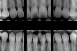
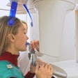
One area that practitioners are always looking to perfect is imaging. Sometimes, little tips and tricks can lead to better images on the first take. Other times, a software update can unlock new features that provide an added layer of confidence when diagnosing. Narrowing in on common imaging problems -- from disinfection to how to maximize your system when offering new treatment -- can create small efficiencies in your workflow that will lead to greater overall productivity as you reopen your practice.
Let's look at some common problems and solutions when it comes to your practice's imaging.
Problem: Patients are extra sensitive to hygiene protocols given the current health crisis and are hesitant to have anything in or near their mouths, including the bite stick of your imaging system.
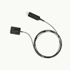
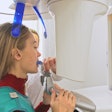
Solution: Disposable panoramic bite sticks can help ease patients' minds about returning to your dental practice for pending dental procedures. Opening and installing a new bite stick in front of the patient can demonstrate your dedication to his or her safety and health. Additionally, using a new stick every time reduces time spent on equipment management as well as lowers costs on materials used for the decontamination and sterilization process.
Problem: The rigidity of your extraoral imaging system forces the patient into an uncomfortable and uncompromising position; minor flaws in positioning can lead to major flaws in the image.
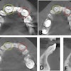
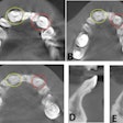
Solution: Minor changes, such as using a different bite block, can often compensate for mispositioning. For example, a Frankfurt block tips the patient's chin down into the ideal position for 3D imaging. For 2D imaging, a new software enhancement called Tomosharp (which you can see an example of below) compensates for less-than-ideal patient positioning by capturing multiple planes in a single acquisition and then analyzing the sharpness of thousands of areas within those slices for a sharper image reconstructed on a 2D plane. Tomosharp is now available with the CS 8100 Evo Edition and CS 8100 3D Evo Edition.
 New Tomosharp technology. Image courtesy of Lea Al Matny, DDS.
Problem: The patient has restorations and implants, causing metal artifacts when taking a 3D image and making it difficult to make a confident diagnosis.
Solution: Typically, visualizing a root fracture line or determining whether an implant is properly integrated can be difficult to do radiographically. This is when software reconstruction algorithms can aid with metal artifact reduction (MAR). For example, CS MAR allows the practitioner to toggle back and forth between the original image and one with the metal artifact reduction filter applied for more confidence when making a diagnosis.
Problem: You want to collaborate on a case with a referral, but you need to ensure all parties have the same view of the case without having to share images by passing along CDs or downloading complicated software with which you (or they) are not familiar.
Solution: First, ensure your software captures DICOM-ready files. This eliminates extra conversion steps, reduces storage, and makes it easier to export to referrals or share with a third party. Next, look for a system that lets you share a full version of your imaging software -- not just a viewer -- to eliminate miscommunication.
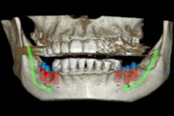
Problem: As a general practitioner, you're branching into the rewarding world of implants. You have been capturing images for years, but getting the perfect image for implant planning and guided surgery is a little different.
Solution: Have the patient sit up tall, secure the head, separate the teeth and cheeks with cotton rolls, and keep the tongue up to separate it from gingiva. Doing so will give you a clearer image of bone for measuring quality and quantity.
Practitioners are problem-solvers, and today the world is facing its biggest healthcare problem yet. Looking at a few of the typical "problems" associated with extraoral imaging, whether it's as simple as repositioning a patient or looking for opportunities to strengthen relationships with referrals, is one way to work smarter in this new environment.
In a world where getting patients in and out of the practice safely and efficiently, what was once mundane -- capturing images with an extraoral system -- has now become a new battleground for infection control, saving time, reducing retakes, and providing more confident diagnoses.
Lea Al Matny, DDS, is a licensed oral and maxillofacial radiologist at SeeThru Reports and clinical education expert at Carestream Dental. She attended the University of Texas Health San Antonio (UTHSA) where she received both a certificate in oral and maxillofacial radiology and a master's degree in dental science. She is a reviewer for Oral Surgery, Oral Medicine, Oral Pathology and Oral Radiology (OOOO) and is actively involved in multiple committees at the American Academy of Oral and Maxillofacial Radiology.
The comments and observations expressed herein do not necessarily reflect the opinions of DrBicuspid.com, nor should they be construed as an endorsement or admonishment of any particular idea, vendor, or organization.
