Calcivis, a medical devices company that is focused on managing tooth decay, announced that it has completed a first clinical study of its Calcivis caries activity imaging system.
The study was the first to evaluate the system in a full clinical dental setting. It was conducted in 39 patients by four general dental practitioners based in three practices in Scotland. Full results and analysis will be completed early in 2015.
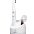
The Calcivis system is an in-clinic device that involves a unique, proprietary bioluminescence approach combined with a specialized imaging device that allows accurate detection and visualization of demineralization by imaging free calcium ions at the tooth surface; a caries lesion that is actively demineralizing is more likely to progress and lead to cavitation, according to the company. The system has already been granted the CE Mark in Europe.
The system combines a sensitive intraoral camera and application technology to deliver a precise amount of disclosing solution, containing a photoprotein, onto the tooth surface. The photoprotein binds calcium ions and emits a blue light signal proportional to the amount of calcium present. This sensitive chemiluminescent system produces a demineralization map of the tooth.
The resulting images provide a focus for discussion with patients about their caries management program and the development of a tailored, rational, evidence-based treatment in line with dental best practice.
"The images have the potential to provide real insights into the ongoing demineralization disease process," Charles Ormond, principal investigator of the study, stated in a press release.

