Cone-beam computed tomography (CBCT) and two injection needles used as reference points assisted in the removal of a broken 1.3-cm suture needle from a man's buccal mucosa, according to a case report published recently in Maxillofacial Plastic and Reconstructive Surgery.
After a dentist accidentally cut the suture needle during the patient's procedure, the clinician tried to use a panoramic x-ray and CT to visualize and retrieve the needle but failed. The dentist was forced to refer the patient to oral and maxillofacial surgeons to have the needle removed, the authors wrote.
"This study reported a rare clinical case of removing the suture needle with the help of CBCT images," wrote the group, led by Dr. Suyun Seon from Kyung Hee University School of Dentistry in Seoul, South Korea (Maxillofac Plast Reconstr Surg, July 5, 2021).
 Photo of the patient's mouth immediately after having explantation and flap surgeries at a dental clinic. All images courtesy of Seon et al. Licensed under CC BY-NC-ND 4.0.
Accidents happen
Breakage of suture needles is a rare but potentially serious occurrence in dentistry. Numerous factors, including limited visibility, difficult access, sudden patient movements, and mishandling of instruments by clinicians, can result in suture materials being lost, broken, or embedded in a patient's oral cavity.
When this occurs, it is vital that foreign objects be removed immediately to avoid serious complications, including massive bleeding and nerve injuries. Imaging helps in the retrieval of foreign bodies, but some imaging technologies work better than others depending on the object and where it is located, according to the authors.
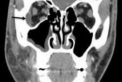
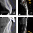
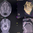
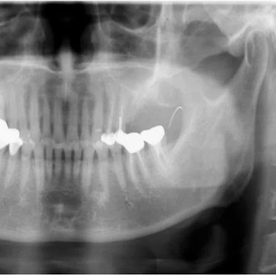
 (A) Panoramic radiograph reveals needle fragment located at the anterior ramus of the left mandible. (B) A CBCT image of the lost needle, with two injection needles used as reference points.
In this case, the man's dentist referred him to the maxillofacial and oral surgery department to have a suture needle removed from his left buccal mucosa. The needle was supposed to be tied with nylon, but the dentist cut it while performing explantation and local flap surgeries due to the man's chronic peri-implantitis, they wrote.
When he arrived at the surgery department, the man had sharp pain at his surgical site, but it did not affect his ability to open his mouth. An x-ray confirmed that the suture needle remained in his left buccal space.
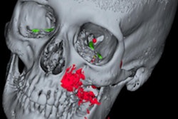
During an oral exam, a surgeon inserted two needles perpendicularly into the man's left buccal mucosa as reference points to better locate the lost suture needle. Then, the patient underwent a CBCT scan that revealed the broken needle was located above the injection needle and below the maxillary tuberosity, the authors wrote.
To remove the broken needle, surgeons opted to place the patient under local anesthesia and conduct a surgical exploration. A vertical incision was made on the man's left buccal mucosa along the ramus, and dissecting scissors were used to make a blunt dissection. However, surgeons stopped the procedure because they couldn't see the needle, and the man was experiencing pain.
After consulting with the patient, they placed him under general anesthesia to remove the broken needle. A deep dissection was made and the 1.3-cm suture needle, as well as the nylon fragment that was attached to it, were retrieved. Then, the incision was closed.
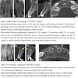
 (A) Photo of single needle fragment lodged in the buccal mucosa. (B) The retrieved suture needle.
After the retrieval, x-rays were taken, which confirmed that the broken needle had been removed completely. One week after the removal procedure, the stitches were removed. The patient was healing and experienced no complications.
Exact localizations and surgical approaches are necessary for removing foreign objects in the head and neck area. CT scans may not measure accurately the real position of a foreign object due to intraoperative traction and swelling, Seon and colleagues wrote.
"Therefore, other up-to-date radiographic devices can be sometimes advisable when CT scan images are not practical," they concluded.

