A treatment plan is usually based on clinical experience, clinical information, and information obtained from diagnostic imaging. But when it comes to endodontic treatment planning, especially for a difficult case, do the images from a cone-beam CT (CBCT) scan actually influence how a specialist proceeds?
The answer from researchers in Spain is yes -- CBCT scans can change the decisions specialists make in endodontic cases. Their study was published in the current issue of the Journal of Endodontics (February 2017, Vol. 43:2, pp. 194-199).
"Our findings suggest that more diagnostic information can be obtained from a preoperative CBCT image than from a preoperative [periapical] radiograph, and that this information can directly influence a clinician's treatment plan, especially in high-difficulty cases," lead study author Gustavo Rodríguez, DDS, and co-authors wrote.
Dr. Rodríguez is on the faculty of dentistry in the department of restorative dentistry and endodontics at the International University of Catalonia in Barcelona.
2 imaging modalities
The decision whether to attempt to save a tooth or extract it is made using an evidence-based approach that includes the interpretation of radiographic images. While periapical radiography (PA) is often used, it only offers a two-dimensional view, while CBCT offers a three-dimensional view.
Other studies have found that endodontists change their treatment plan more than 60% of the time after viewing CBCT images, but no study has found if this is also true among dental specialists, such as oral surgeons, periodontists, and prosthodontists, especially when confronted with a difficult case, the study authors noted.
So the researchers of the current study wanted to see if specialists did change their treatment plans after viewing preoperative CBCT images of patients with varying degrees of treatment complexity.
“More diagnostic information can be obtained from a preoperative CBCT image than from a preoperative [periapical] radiograph.”
The study included 100 specialists, including the following:
- 40 endodontists
- 40 prosthodontists
- 32 periodontists
- 28 oral surgeons
The researchers chose 30 cases of differing levels of endodontic difficulty: 10 each of minimum, moderate, and high difficulty. They sought to simulate a patient's first visit to a specialist by including at least two clinical photographs, two PA radiographs, and a bitewing for the posterior teeth for each case. The case also included the patient's clinical history.
The 30 cases were presented to all 100 specialists at the same time in random order. The specialists were asked to choose one of seven treatment alternatives, which ranged from no treatment necessary to extraction.
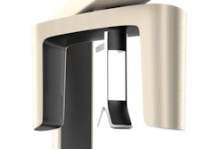
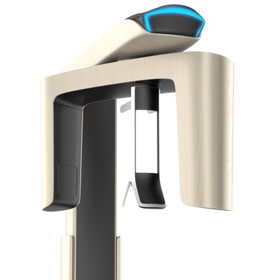
One month later, the practitioners were reassembled to review the information, but this time images from a small-volume CBCT scan (Planmeca ProMax 3D s, Planmeca) were included.
The number of specialists who chose each treatment option changed after the viewing of the CBCT images. The number of specialists who opted for apical surgery, nonsurgical treatment and apical surgery, or extraction increased after viewing the images, while the number who chose no treatment or watchful waiting decreased (see table below). The number of specialists who chose endodontic treatment remained about the same: 2,738 before viewing the CBCT images and 2,664 after.
| Treatment changes before and after CBCT scan | ||
| Treatment alternative | Before CBCT scan | After CBCT scan |
| No treatment | 56 | 22 |
| Watchful waiting | 202 | 108 |
| Apical surgery | 274 | 306 |
| Nonsurgical retreatment and apical surgery | 132 | 198 |
| Extraction | 402 | 632 |
According to the study results, the number of practitioners who chose the extraction option rose considerably. Just under 10% of practitioners chose extraction as the treatment course after the first evaluation, the study authors reported. But after the second examination that included CBCT images, that number rose to more than 15%. This increase was statistically significant in all specialist groups, they noted.
The study also measured the self-reported level of difficulty in making a treatment choice both the first examination and after the second examination. The specialists found that the level of difficulty in making a decision increased after the second examination (see table below).
| Percentage change in treatment decision difficulty | |||
| Decision difficulty | 1st exam | 2nd exam | Percentage change |
| Easy decision | 52.1% | 43.67% | -8.43% |
| Moderate decision | 30.14% | 35.62% | +5.48% |
| Hard decision | 17.76% | 20.71% | +2.95% |
"All groups reported a relatively low decision-making level of difficulty when viewing the information using PA radiographs. However, this trend reversed after the CBCT evaluation, increasing the difficulty of the decision-making level," the authors wrote.
Complicated process
The authors noted that results concerning the level of difficulty in making a treatment decision cannot be compared with other studies because of the lack of similar research. In addition, practitioners should be mindful of the radiation dosage to which patients are exposed, they wrote. In this study, the Promax 3D s system was used because it minimized radiation exposure.
However, preoperative CBCT offers more information to a practitioner, even if it complicates the decision-making process, they noted.
"This leads us to hypothesize that a preoperative CBCT image provides more diagnostic information, even though this information is not always easy to assess," the authors concluded.
