Correctly identifying the anatomy of a root canal is crucial for successful endodontic therapy. Researchers tested two imaging modalities to see if either was more accurate than a gold standard.
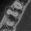
Cone-beam CT (CBCT) performed better than periapical radiography in detecting the apical delta in more than 100 premolars, but both modalities fell short compared with the gold standard of microcomputed tomography (micro-CT), they reported in a study published in the Journal of Endodontics (May 2019, Vol. 45:5, pp. 549-553).
CBCT imaging showed better performance than periapical radiography in the detection of apical deltas, wrote the authors, led by Eduarda Helena Leandro, DDS, PhD, of the department of oral diagnosis at the University of Campinas Piracicaba Dental School in Sáo Paulo, Brazil.
Anatomic complexities
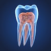
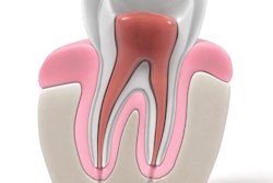
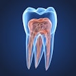
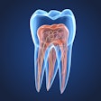
Endodontic treatment is often challenging because of anatomic complexities, such as apical deltas in which the main root canal is divided into multiple ramifications in the apical third. Misdiagnosis may lead to treatment failure and additional complications. Knowing which imaging method is more accurate in detecting apical deltas influences clinical decision-making.
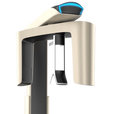
The researchers took images of 110 human premolars with 120 root canals. They used the VistaScan digital intraoral system (Durr Dental) for the periapical radiographic images and the 3D Accuitomo CBCT unit (J. Morita) for the CBCT images. They also took images of the canals with a micro-CT system to serve as the gold standard for the study.
Two oral radiologists assessed the images for the presence of apical deltas using a five-point scale. Additionally, the number of apical foramina also was evaluated in the CBCT images.
Apical deltas were present in 40 root canals according to the micro-CT images, the researchers reported. They found that the periapical radiographs and CBCT images differed significantly from the gold standard (p < 0.05) in detecting apical deltas (see table below).
| Comparison of diagnostic test results for CBCT and periapical radiography in identifying apical deltas | ||
| CBCT | Periapical radiography |
|
| Accuracy | 0.73 | 0.62 |
| Sensitivity | 0.35 | 0.07 |
| Specificity | 0.92 | 0.90 |
| Positive predictive value | 0.70 | 0.27 |
| Negative predictive value | 0.74 | 0.66 |
CBCT imaging showed higher values than periapical radiographs for all diagnostic tests, the researchers reported. However, both modalities had very low sensitivity values.
CBCT images also tended to underestimate the number of foramina present (p < 0.05), they added.
"Despite its moderate accuracy and high specificity, CBCT showed low sensitivity, which means that many positive cases were not detected by the observers," the authors noted.
Critical approach
While the researchers listed no study limitations, they stated that the presence of root canal filling could reduce accuracy and recommended further studies on this subject.
While previous studies have considered CBCT as a gold standard, the study authors recommended a more critical approach to the modality's use.
"In general, the number of apical foramina cannot be reliably assessed using CBCT imaging," they concluded.

