A team of U.S. researchers will use genetically engineered mice and 3D imaging technology to study the development of the human midface -- upper jaw, cheekbones, and eye sockets -- and how diseases and abnormalities of the head affect the growth and shape of the face.
The work is being funded by a $2.3 million five-year grant from the National Institute of Dental and Craniofacial Research.
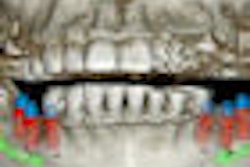
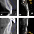
The researchers will measure facial tissues and spaces, using specialized 3D images to learn more about these defects in human patients, and will use genetically engineered mouse models to guide investigations of these human diseases.
"These combined approaches will lead to the discovery of the underlying molecular, cellular and developmental basis of these disorders, which include the craniosynostosis syndromes, like Apert and Crouzon syndromes," said lead researcher Joan Richtsmeier, PhD, a professor of anthropology at Pennsylvania State University, in a press release. "We hope our work will eventually lead to the improvement of care for patients with these conditions."
Craniosynostosis is a condition in which the fibrous tissues uniting the bones of the skull close too early, causing problems with normal brain and skull growth. Premature closure of these tissues surrounding the brain is often associated with changes in facial bones, commonly called midface hypoplasia. Many diseases of the head share the common feature of midface hypoplasia, a medical condition in which the person's upper jaw, cheekbones, and eye sockets are smaller than normal.

