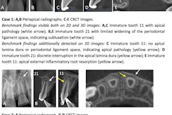
While cone-beam CT (CBCT) provides more information than traditional radiography about impacted and supernumerary teeth in pediatric patients, practitioners should consider the radiation risks when choosing which diagnostic method to use, according to a study in Pediatric Dentistry (July-August 2010, Vol. 32:4, pp. 304-309).
Researchers from the University of Texas Health Science Center at Houston surveyed 10 pediatric dental faculty and 10 pediatric dental residents after they had viewed eight clinical cases in either cone-beam CT or traditional radiography in which the patient presented with pathology in the anterior maxilla. The surveys asked for pathology diagnosis, location, and identification of root resorption, as well as questions about the usefulness of the radiographic mode in treatment planning.
The researchers found that cone-beam CT was statistically better (p < 0.05) for pathology location, determining root resorption, and adequacy in treatment planning. More faculty members were able to correctly identify the pathology location (p = 0.034), while more residents believed they could determine presence of root resorption (p = 0.029). For impacted versus supernumerary cases, more pathology was correctly located when viewed using cone-beam CT (p < 0.05).
Although cone-beam CT provides more information than traditional radiography on the location of pathology, the presence of root resorption, and treatment planning, "the benefits of CBCT imaging must be weighed against the radiation risk to the pediatric patient and the complexity of the pathology," the researchers concluded.
Copyright © 2010 DrBicuspid.com


