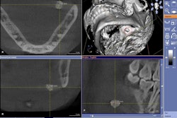
Cone-beam CT (CBCT) can measure distances from the apices of selected posterior teeth to the mandibular canal as accurately as direct anatomic dissection, according to a study in the Journal of Endodontics (July 2010, Vol. 36:7, pp. 1191-1194).
Researchers from the Loma Linda University School of Dentistry conducted a pilot study to determine the scanning parameters that produced the best diagnostic image and the best dissection technique. Using an I-CAT Classic cone-beam CT system (Imaging Sciences International), they scanned 12 human hemimandibles with posterior teeth and exported the images into DICOM format.
The researchers examined the scans using InVivo Dental software (Anatomage) and measured the apex of each root along its long axis to the upper portion of the mandibular canal. The specimens were dissected under a dental operating microscope, and analogous direct measurements were taken with a Boley gauge. All measurements were taken in triplicate at least one week apart by one individual.
The researchers found no statistical difference between the methods of measurement (p = 0.676). Both measurement methods were highly predictive of and highly correlated to one another according to regression and correlation analysis, they noted.
"Based on the results of this study, the I-CAT Classic can be used to measure distances from the apices of the posterior teeth to the mandibular canal as accurately as direct anatomic dissection," the researchers concluded.
Copyright © 2010 DrBicuspid.com


