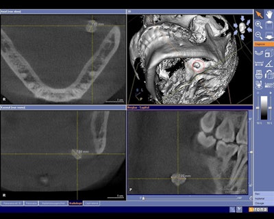
 Cone-beam CT dataset showing a slightly overaverage size salivary calculus of 7-8 mm in the left submandibular gland area in the axial (upper left), coronal (lower left), and sagittal (lower right) view. Maximal diameters were measures using the visualization software tools. In the 3D reconstruction (upper right), the reformatted 3D salivary calculus is visualized and marked with a red circle. All images courtesy of Timo Dreiseidler, M.D., D.M.D., University of Cologne. |
 Cone-beam CT dataset showing a 2-mm salivary calculus in the left submandibular gland area in the axial (upper left), coronal (lower left), and sagittal (lower right) view. Clinical and conventional 2D diagnosis failed here due to the small size of the calculus. |
| close |