Should dental schools consider teaching dental students how to evaluate patient x-rays for signs of osteoporosis?
Yes, according to a study in the Journal of Dental Education (May 1, 2013, Vol. 77:5, pp. 598-603).
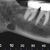
A clinician from the College of Dentistry at the University of Tennessee Health Science Center selected 25 panoramic radiographs of dental school patients with a history of osteoporosis and radiographic changes suggestive of the disease and 25 normal panoramic radiographs.
Twenty dental students were then taught to use the mandibular cortical index to detect changes suggestive of osteoporosis. The students also used a five-point scale to determine the diagnostic accuracy of panoramic images for osteoporosis. Their diagnoses were compared to the gold standard -- a dental clinician -- and an oral radiologist.
The interrater consistency was excellent for both the students and between the students and the radiologist, the study authors reported, and there was no statistically significant difference between radiographic and clinical evaluations (p = 0.0801).
"Teaching dental students to recognize radiographic changes suggestive of osteoporosis in routine panoramic radiographs should be emphasized to improve their awareness and identification of this disease," the researchers concluded.