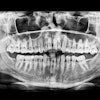
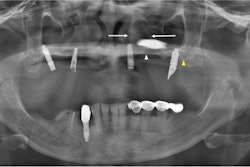
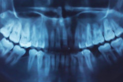
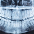
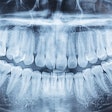
Panoramic x-rays can help practitioners better predict maxillary canine impaction, according to a new study in the American Journal of Orthodontics and Dentofacial Orthopedics (July 2012, Vol. 142:1, pp. 45-51).
Treatment of impacted maxillary canines frequently requires surgical intervention, which can involve substantial complications, noted the authors, led by Anand Sajnani from Terna Dental College and Hospital in Mumbai, India.
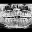
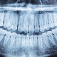
The retrospective study was conducted at a dental hospital in Hong Kong to determine whether impaction of a maxillary canine can be predicted using measurements made on a panoramic radiograph. Geometric measurements were made on 384 panoramic radiographs of patients with a unilaterally impacted maxillary canine (group 1) to characterize its presentation and compare them with the unaffected antimere (group 2).
In patients 8 years and older, the researchers found a "clinically discernible difference" of 4 mm between the mean distance of the tip of the impacted canine (group 1) and that of the antimere (group 2) from the occlusal plane (p < 0.05). They also found a statistically significant difference at the age of 9 years and beyond between the two groups according to the position in different sectors and according to the mean angle made with the midline (p < 0.05).
"Diagnosis of maxillary canine impaction is possible at 8 years of age by using geometric measurements on panoramic radiographs," the study authors concluded.


