Dear Imaging & CAD/CAM Insider,
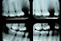
The American Academy of Oral and Maxillofacial Radiology (AAOMR) has published a new position statement regarding the use of imaging in implantology -- the first update of these guidelines since 2000.
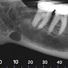
Given the increasing adoption of cone-beam CT for dental implant planning over the last decade, the revised guidelines put particular emphasis on this modality.
Click here to read the AAOMR's new recommendations in this Imaging & CAD/CAM Insider Exclusive.
In related news, some Oregon dentists are reportedly opting not to treat patients who refuse to be x-rayed before having their teeth cleaned. The Oregon dental board recommends dentists follow ADA and U.S. Food and Drug Administration (FDA) guidelines to ensure they are meeting the standard of care with regard to radiographs. Read more.
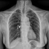
And the FDA has raised the ante in its drive to reduce pediatric radiation dose, launching a new program that will require manufacturers to consider the safety of children when designing new x-ray imaging devices. Devices that do not comply with the program may have to carry warning labels advising against their use in children.
In other imaging news, people who received frequent dental x-rays appear to have an increased risk of developing meningioma, according to the largest case-control study to date examining the correlation between dental x-rays and this largely benign brain tumor. But several members of the imaging community found major flaws in the study, including the AAOMR.
Meanwhile, independent practice dental hygienists in Maine are claiming victory with the passage of legislation that allows them to take a broad range of x-rays in federally designated underserved areas of the state as part of a pilot project designed to address the state's severe access-to-care issues. Read more.
On the technology front, when compared to a high-end, consumer-grade liquid crystal display monitor, the iPad 2 fared well in the display of in vivo images for the assessment of interproximal caries, according to a study published in Oral Surgery, Oral Medicine, Oral Pathology, Oral Radiology.
And researchers from the University of Texas at Austin are turning the high-resolution imaging capabilities of confocal microscopy into an affordable and user-friendly clinical tool for detecting oral cancer and enabling real-time assessment of surgical margins, while a bioengineering professor at Stanford University has developed a low-cost way to use smartphones to create images of the oral cavity and screen patients' mouths for suspicious lesions.