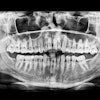
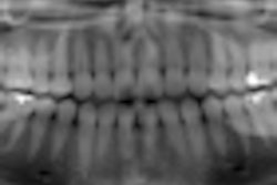
Dental radiography is often used for detecting marginal gaps at the implant-abutment interface, but the angulation of the x-ray beam may lead to inaccurate diagnosis in gap detection, according to researchers from the University of Athens (Journal of Esthetic and Restorative Dentistry, August 2010, Vol. 22:4, pp. 235-250).
To investigate the accuracy of dental radiography in detecting marginal gaps at the implant-abutment interface, the researchers took radiographs on internal and external hex implants with different experimental gaps and inclinations.
The abutment was screwed on the implant, and the implant was placed into a box filled with silicone impression material. The x-ray film was placed parallel to the implant at the back of the box, and the borders of the box were marked to the base and the box.
A 10-cm ruler was attached to a long x-ray tube to ensure parallelism to the implant. Sets of radiographs were made at 0°, 5°, 10°, 15°, 20°, 25°, and 30° (to the abutment) and −5°, −10°, −15°, −20°, −25°, and −30° (to the implant).
After observing the images with visual examination, under magnification, and in a slide projector, the researchers noted that the ability to diagnose a gap at the implant-abutment interface varied significantly with the angulation degree of the x-ray tube. In all examinations, the gap was not detectable at angulations higher than 20°, while in visual examination at 25° and 30°, an average clinician was able to diagnose the distortion.
"The x-ray diagnosis of gap at the interface can be significantly influenced by the inclination of the x-ray tube in relation to the long axis of the implant," the researchers noted. To achieve accurate results, the use of a paralleling device is advocated to achieve greater detection ability, they concluded.
Copyright © 2010 DrBicuspid.com