What effect does explaining a preoperative radiograph prior to third-molar surgery have on patient satisfaction with understanding potential complications and risks of the surgery? Not much, according to a new study in Dentomaxillofacial Radiology (March 2010, Vol. 39:3, pp.176-178).
Researchers from Aarhus University School of Dentistry set out to compare patient satisfaction with the preoperative information before and after explaining the radiograph, and compare patient satisfaction with the radiographic information when based on a digital or a conventional image and on extraoral or intraoral images.
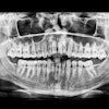
They provided 263 patients (with 301 mandibular third molars) with preoperative information before removal of the third molar. The course of the surgery and possible postoperative complications and risks were explained to the patient. The patients rated their satisfaction with the information on a 100-mm visual analogue scale (VAS). The radiograph was displayed and the radiographic information was explained to the patient, with emphasis on tooth-specific risk factors. The patients again rated their satisfaction on a VAS.
The researchers found no significant difference in patient satisfaction score before (mean VAS = 92.5 mm) and after (mean VAS = 91.7 mm) the radiographic information was explained (p = 0.15). The type of image shown had no bearing either, they found; no difference in satisfaction was found between patients who were shown either digital or conventional images, or between patients who were shown either extraoral or intraoral images (p > 0.5).
"No additional patient satisfaction was obtained by showing and explaining the radiograph to the patient before lower third-molar surgery," the authors concluded. "If the dentist still wishes to show the patient the radiograph, the type of image seems not to be important."
Copyright © 2010 DrBicuspid.com