Editor's note: Edwin T. Parks' column, Talking Pictures, appears regularly on the DrBicuspid.com advice and opinion page, Second Opinion.
Once you've entered the world of three dimensional imaging, it's hard to go back to 2D.
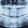
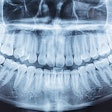
For years I've interpreted countless periapical and panoramic images, feeling quite comfortable that my assessment of three dimensional structures flattened into two dimensions was adequate. All of us grew up using 2D imaging and for most of what we do as clinicians, 2D imaging is fine. But now that I know what 3D imaging can provide, it's sometimes hard to base a clinical decision on 2D imaging.
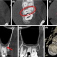
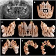
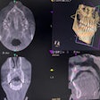
Obviously, I'm not talking about caries or periodontal disease. Usually, the issue is about the location of one object as it relates to other objects, such as the positioning of an impacted canine, the relationship of the mandibular canal space and an unerupted third molar, or the buccal-lingual location of a soft-tissue calcification. After using 3D imaging it becomes apparent that a lot of those object relationship issues were determined with less than optimum data. Why rely on two periapicals and the SLOB rule or a panoramic image and an occlusal projection when you can look at the object of interest in multiple images with multiple orientations?
Three-dimensional imaging has helped to significantly diminish the number of times I use the terms "probably" or "most likely" when communicating about radiographic findings.
I must disclose, however, that the above-mentioned terms are still used when communicating to my teenage children. But at least it's a parental waffle as opposed to a radiologist's waffle!
The comments and observations expressed herein do not necessarily reflect the opinions of DrBicuspid.com, nor should they be construed as an endorsement or admonishment of any particular idea, vendor, or organization.
Copyright © 2008 DrBicuspid.com
